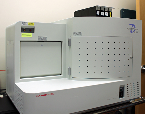
The Digital Imaging room in the Pathology Department at VCU Health. The Hammamatsu NanoZoomer 2.0 HT scanner is essential for digitizing and archiving cytopathology specimens.
The Virginia Commonwealth University Department of Pathology's efforts to provide the highest level of care combined with the challenge of managing a growing archive of over 20 years of glass slides has motivated the department to embrace integrating digital imaging solutions into the clinical and research workflows. Digital pathology allows for rapid access to prior case materials and easy access to extensive research materials.
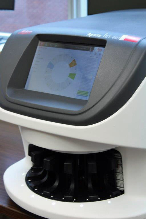
The Leica Aperio GT450 digital Imaging scanner at is capable of holding 450 slides and scanning those slides at an average time of 32 seconds for an output of 81 slides per hour at 40x magnification.
Currently, the division of anatomic pathology has two Leica Aperio GT450 scanners and a Hammamatsu NanoZoomer 2.0 HT scanner. Each GT450 is capable of holding 450 slides and scanning those slides at an average time of 32 seconds for an output of 81 slides per hour at 40x magnification. With the massive throughput afforded by these scanners, we are able to digitize our daily clinical case volume as well slides from our extensive archive. The NanoZoomer scanner is essential for digitizing and archiving cytopathology specimens on multiple focal planes in a process known as Z stacking.
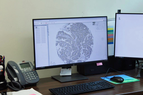
A digital image displays on the screen. VCU pathologists are able to provide intradepartmental consultations from anywhere with Internet access.
The digital imaging platform at VCU is validated for clinical use which allows for case verification across multiple onsite and offsite locations. Additionally, our pathologists are able to provide intradepartmental consultations from anywhere with Internet access. Our busy extradepartmental consultation service has benefited greatly from digital pathology. All outside cases are scanned prior to return providing on-demand access to a patient's prior case for comparison as well as future utilization in research studies. As more pathology departments gain access to digital imaging, the need to send slides through mail will become obsolete.
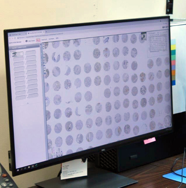
Digital images are displayed on the screen. Digital pathology allows for rapid access to prior case materials and easy access to extensive research materials.
The potential for collaborative research within digital pathology is enormous. Artificial intelligence and machine learning are revolutionizing the pathology landscape by increasing diagnostic accuracy, decreasing pathologist review time, and providing prognostic information that would otherwise not be possible. The use of artificial intelligence in pathology is still relatively new and we are well suited to leveraging our vast archive to contribute to ongoing research efforts.
Leadership and Contacts

Bryce Hatfield, MD
Director of Digital Imaging and Gastrointestinal Pathology; Assistant Professor
Bryce Hatfield, MD
Director of Digital Imaging and Gastrointestinal Pathology; Assistant Professor
Pathology
Director of Surgical Pathology Operations
Phone: (804) 628-2949
Email: Bryce.Hatfield@vcuhealth.org