Renal Pathology Services is open Mon-Fri 8am-5pm; Call Client Services 804-828-PATH (828-7284); 1-800-363-9234
Renal Pathology Services offers expert renal biopsy interpretation and consultation to nephrologists and transplant surgeons. We utilize the most advanced technologies available including light microscopy (LM), electron microscopy (EM), and immunofluorescence (IF).
24 Hour Turnaround & Same-Day Processing
A written, preliminary diagnosis based on LM and IF is offered within 24hrs. Same-day processing is offered for transplant biopsies, prepared slides, and selected tissue samples.
Tests Performed
- LM utilizing H&E, PAS, methenamine silver, Masson trichrome stains, special stains, such as congo red, as indicated
- IF utilizing a full battery of fluorescein-tagged antisera (IgA, IgM, IgG, C3, C1q, kappa, lambda, fibrinogen)
- EM on biopsies as appropriate
Specimen Handling
Client Services 804-828-PATH (828-7284); 1-800-363-9234
! Fresh State
Immediately place the renal tissue core, or open biopsy wedge in a secure container with physiologic saline solution or sterile culture media to prevent drying. Or, alternatively, wrap the specimen in saline-moistened non-stick dressing. Avoid compression and drying of the specimen.
Separation Prior to Transport
If the specimen is divided prior to transportation, the proper fixative solutions for LM, IF, and EM must be available in your institution. A formalin-filled container for LM, Michel's medium for IF, and glutaraldyhyde for EM studies will be supplied upon request.
When dividing the specimen, each part must contain glomeruli!
If an open biopsy, or 3 or more tissue cores are obtained:
- one portion or core is submitted for IF
- one for EM
- the remainder for LM
If 2 tissue cores are obtained:
- one is submitted for LM
- the second is cut into four 1-2mm fragments: two are submitted for EM, and the other two for IF
If one tissue core is obtained and it is small, <8 mm:
- it should be submitted fresh in saline or in a saline-moistened non-stick dressing
- when dividing a small biopsy, note priority order (1 is top priority):
- LM
- IF
- EM
If single core is >8 mm:
- divide it into two portions and submit one half for LM
- divide the remaining half for IF and EM
Special Instructions
A surgical pathology Accession Sheet with adequate clinical history, differential diagnosis, and laboratory findings must accompany the specimens for their proper interpretation.
Please contact Client Services 804-828-PATH (828-7284); 1-800-363-9234 for assistance with the Accession Sheet and detailed information about transportation, specimen handling, separation prior to transportation and causes for rejection.
The Anatomic Pathology Receiving area is located at the MCV campus in the Gateway Building, 1200 E. Marshall Street, 6th floor.
Leadership and Contacts
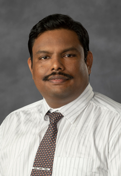
Selvaraj Muthusamy, MBBS, PhD
Renal and Genitourinary Pathology; Assistant Professor
Selvaraj Muthusamy, MBBS, PhD
Renal and Genitourinary Pathology; Assistant Professor
Pathology
Director of Molecular Genetic Pathology Fellowship
Phone: (804) 628-6070
Fax: (804) 828-8733