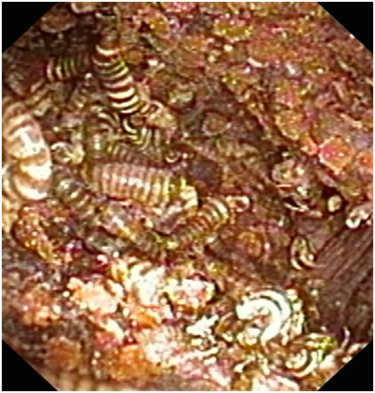
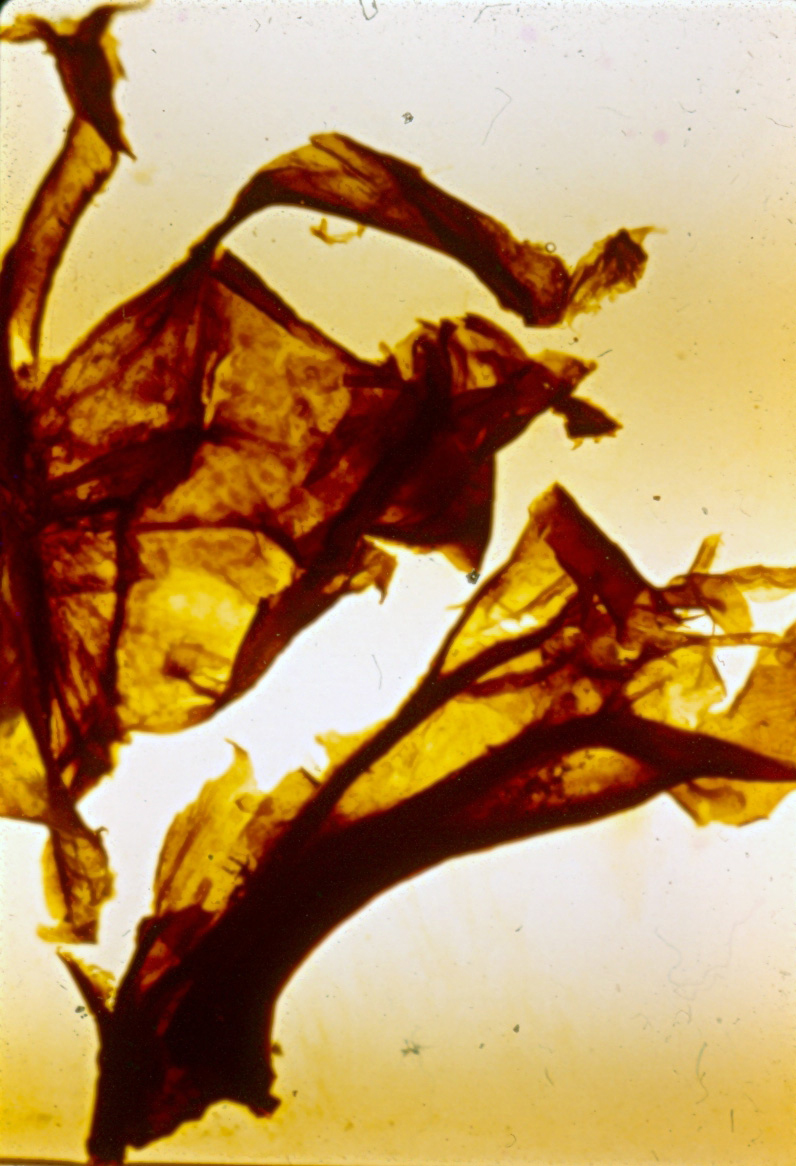
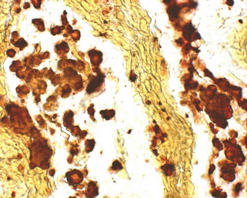
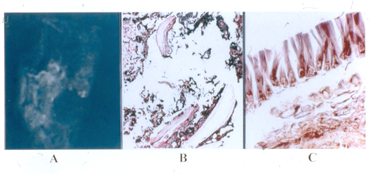
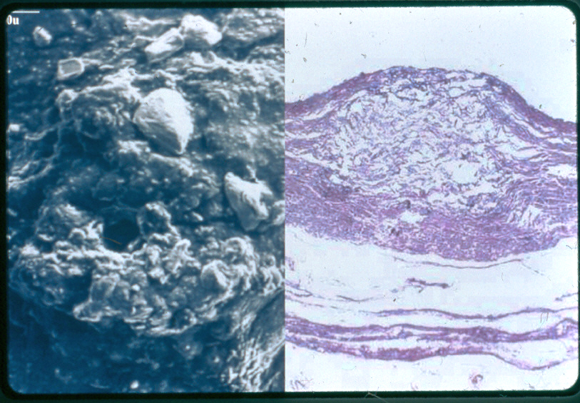
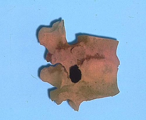
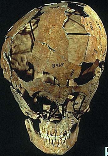
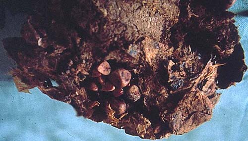
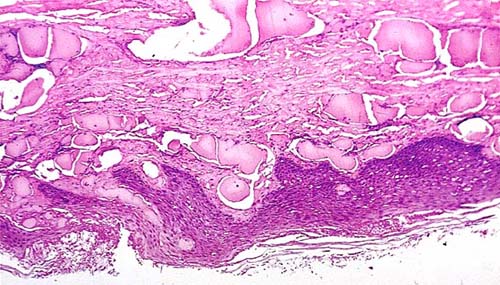
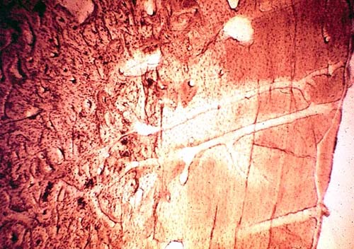
Case 150
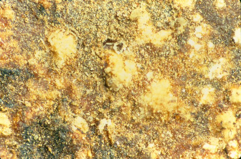
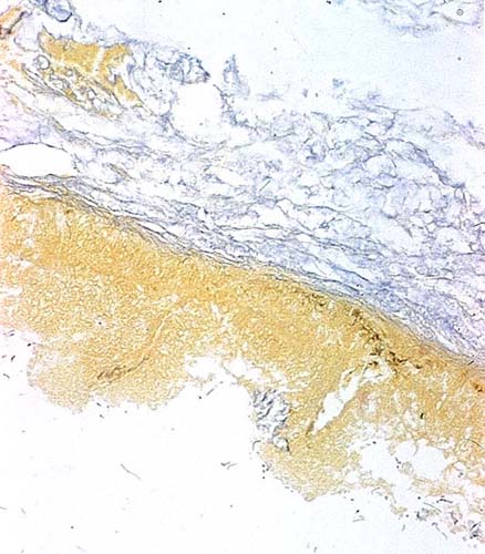
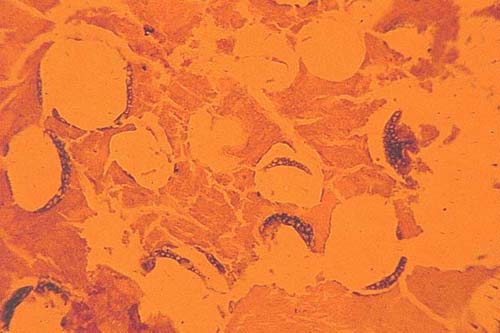
History: This is a montage of some of the unusual particles found in the tissue sections of the lungs of mummies from the Azapa Valley. (~ 1100 BC t0 1000 AD). What are they? What might they signify?
.jpg)
Submitted by: Jerrold L. Abraham, MD, SUNY Upstate Medical University, Syracuse, NY, USA.
Diagnosis: Answer for Case 150 (pdf)
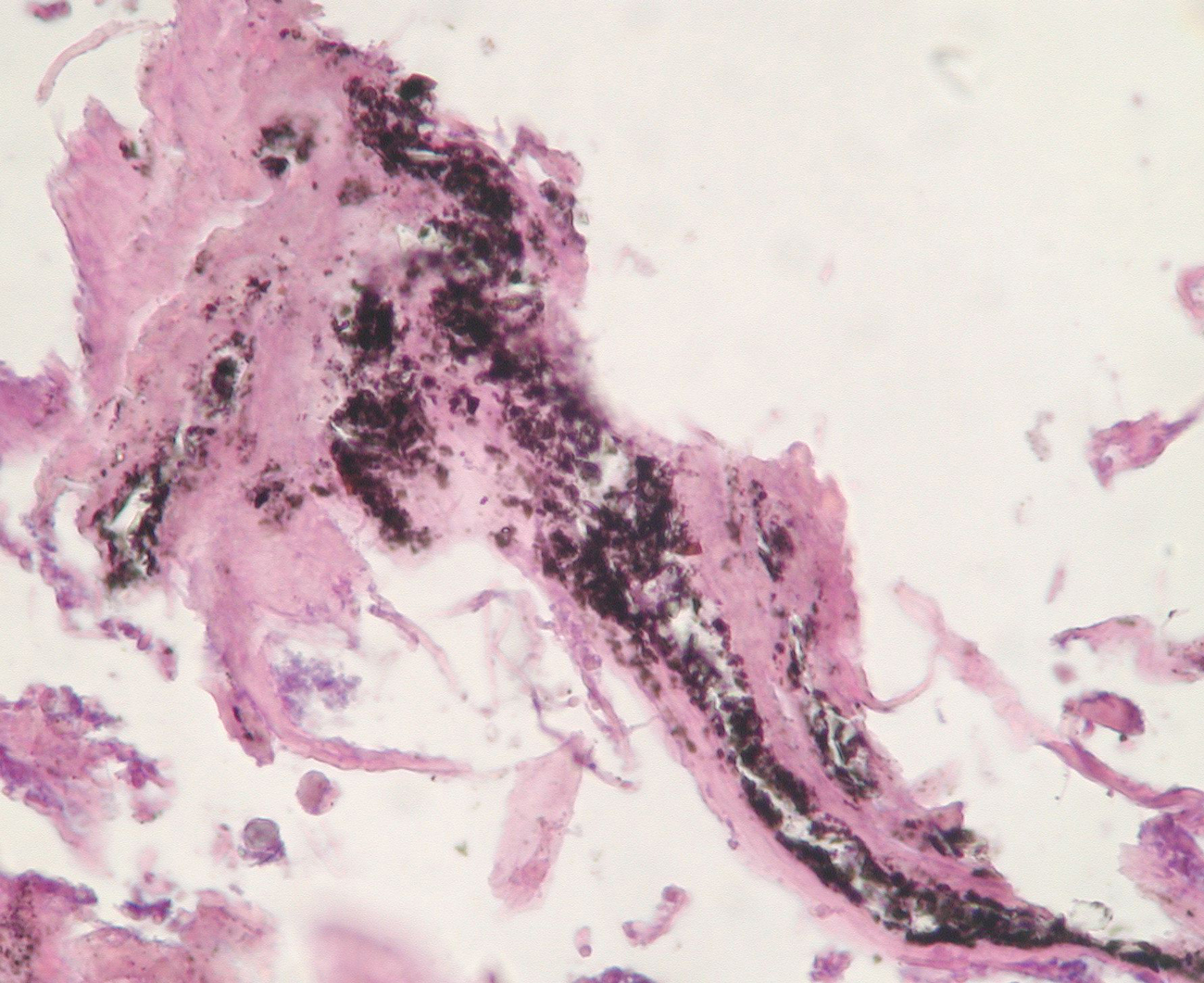
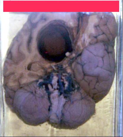
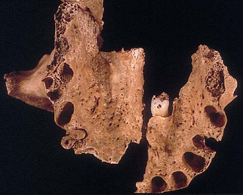
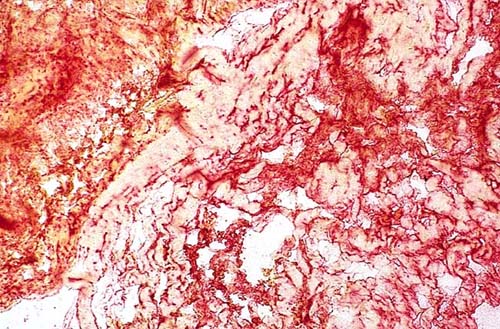
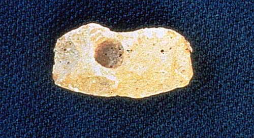
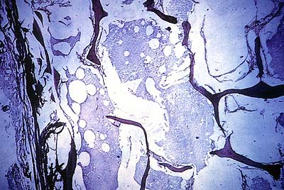
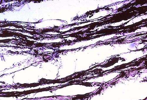
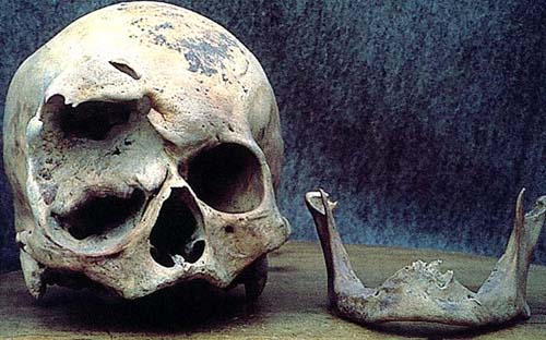
Case 149
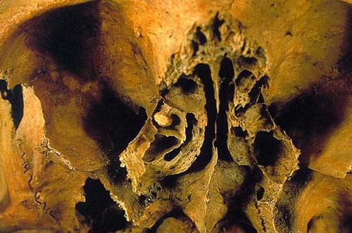
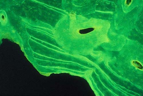
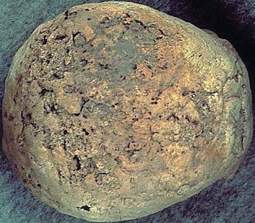
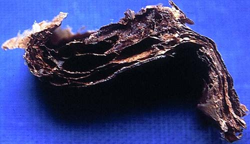
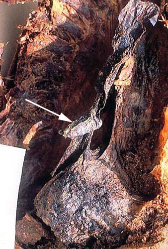
History: Histological section of mummified intracranial material from an individual of the Mediterranean Bronze
Age showing reticular tissue. How do you interpret the structure on the left of this mummified tissue?
Submitted by: Pedro L. Fernández, MD, PhD., Hospital Germans Trias i Pujol, Universidad Autonoma de Barcelona, Department of Pathology, Barcelona, SPAIN.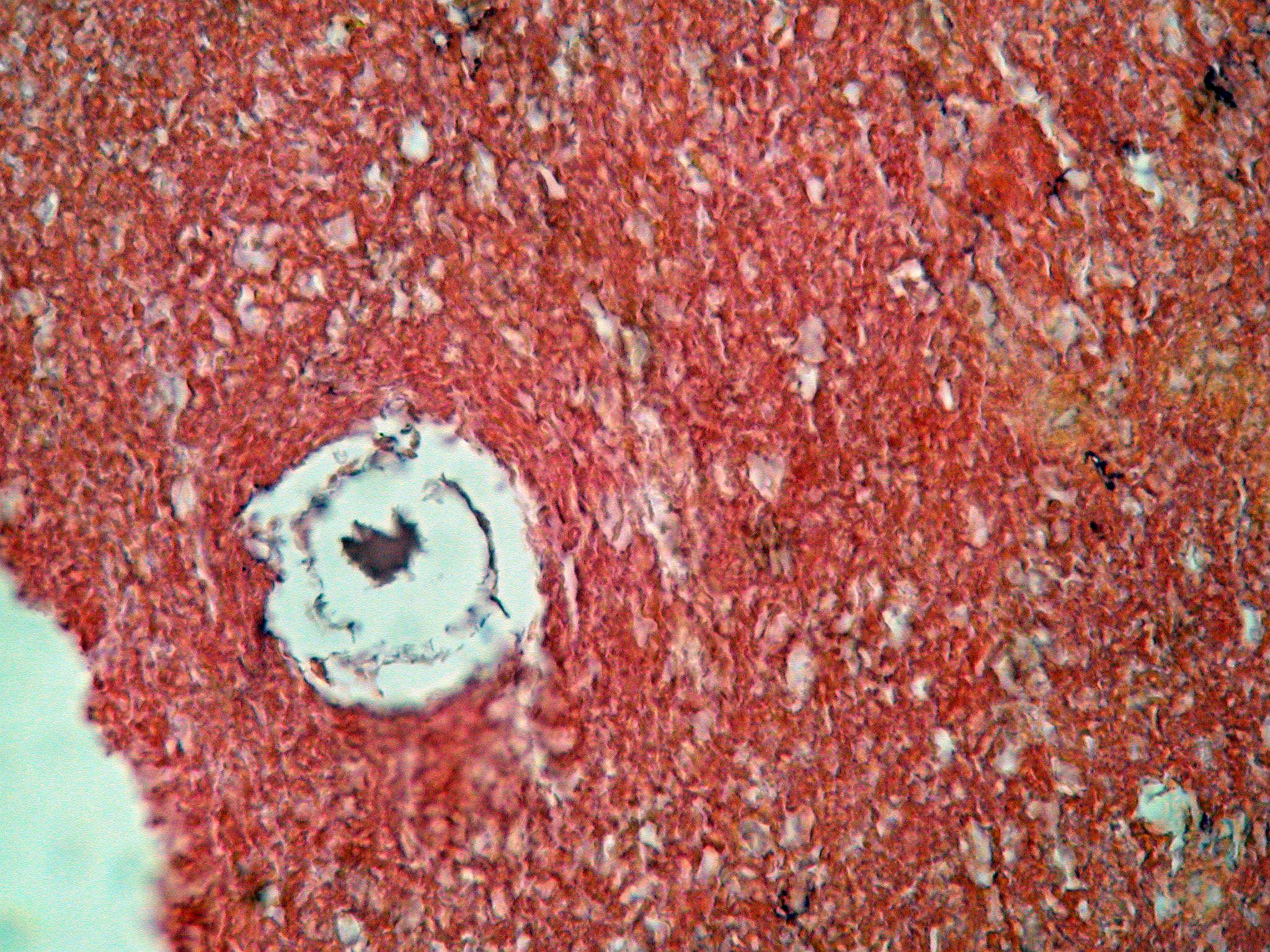
Diagnosis: Case 149 Reticular tissue with a round space consistent with nervous tissue containing a vascular and perivascular space on the left. HE,x 200.
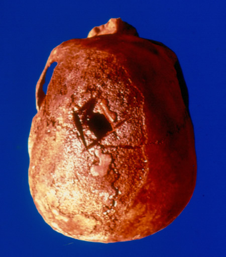
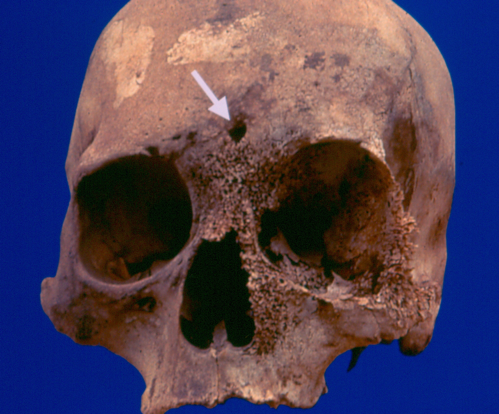
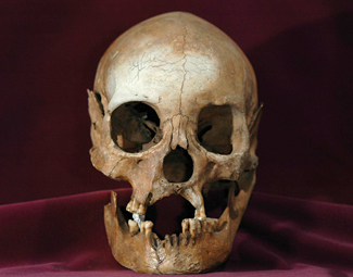
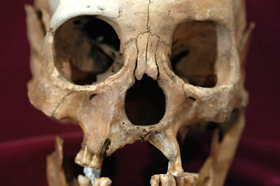
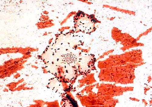
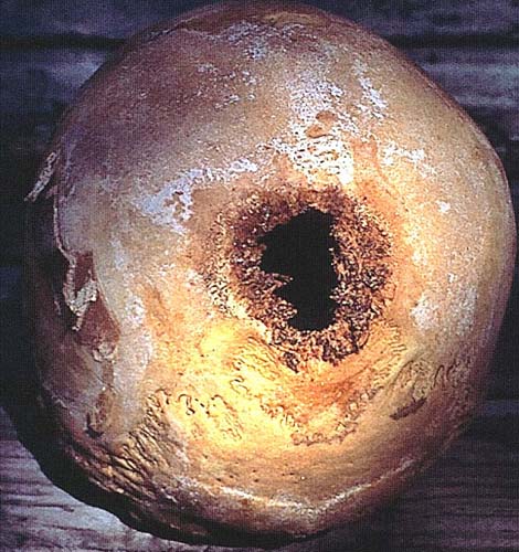
Case 148
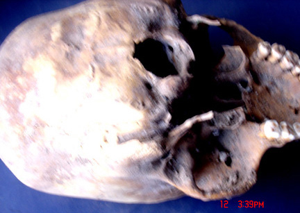
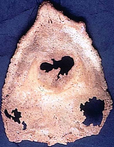
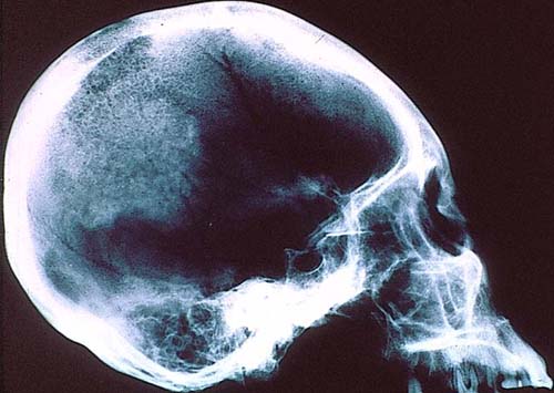
History: Skull from Chanka Culture, Andahuaylas, Peru (ca. AD 1150-1250).
Submitted by: Danielle S. Kurin, University of California, Santa Barbara, CA
Diagnosis: Case 148 Artificial Cranial deformation.
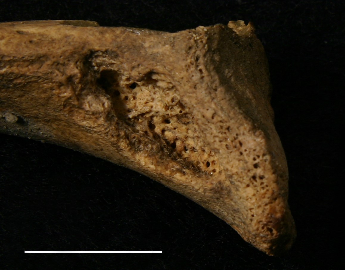
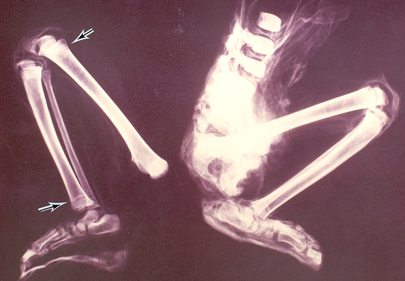
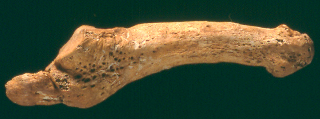
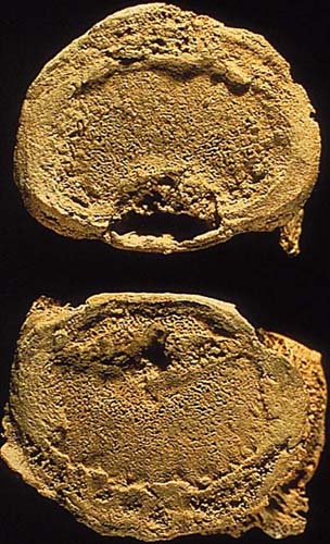
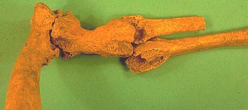
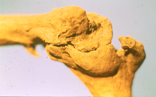
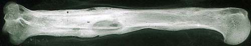
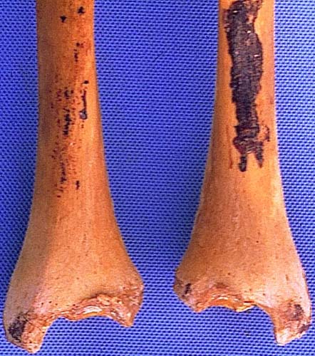
Case 147
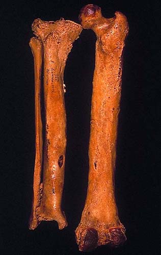
Femur bones of 13th century female aged 45-55 years at time of death, Poggibonsi, Siena, Italy.
Submitted by Dr. Valentina Giuffra, University of Pisa, Italy.
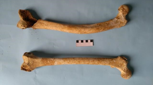
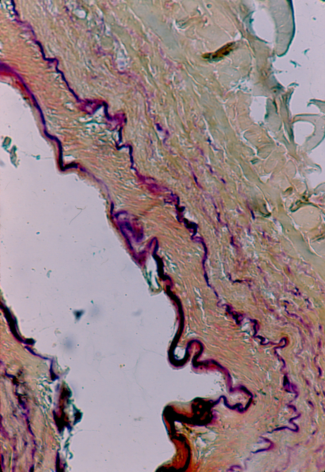
Diagnosis: Case 147 Greenstick fracture, probably occurred during the infantile age (absence of a true callus).
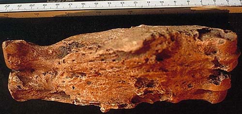
Case 146
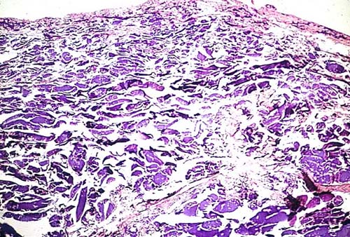
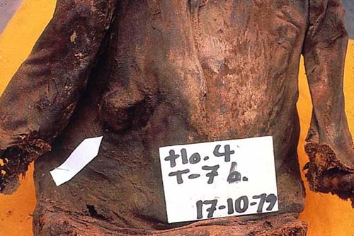
Subcutaneous mass on the right side of the chest from a male aged 14 of Northern Chile from 1,100 to 1,200 AD.
Submitted by Dr. Enrique Gerszten, Virginia Commonwealth University, Richmond, Virginia.
Diagnosis: Case 146 Lipoma. The histology shows conglomeration of fat cells intermingling with fibrous septa.
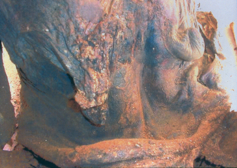
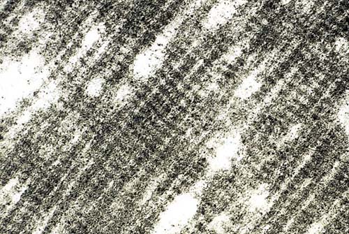
Case 145
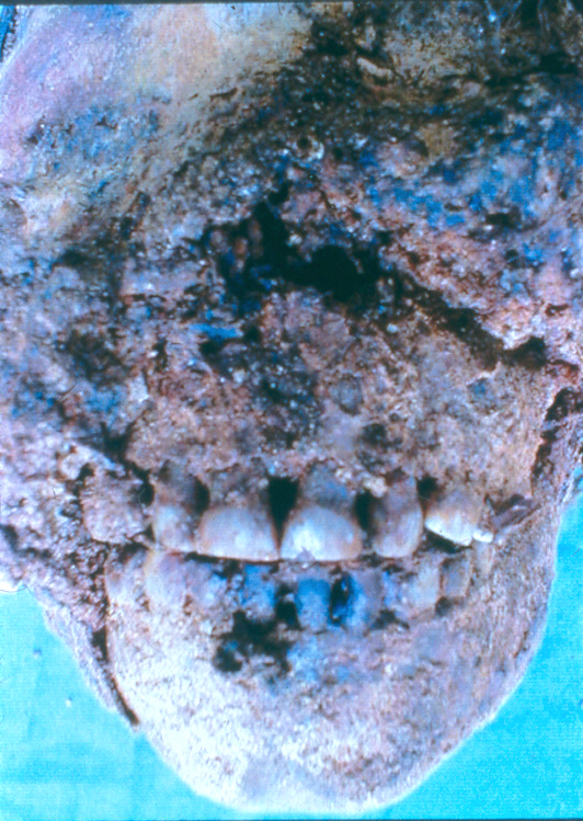
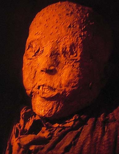
The slide shows the face of a priest from the early XVII Century entombed in the basement of the Church “Iglesia del Carmen” in Mexico City.
Submitted by Dr. Enrique Gerszten, Virginia Commonwealth University, Medical College of Virginia, Richmond, VA.

Diagnosis: Case 145 Smallpox. We don’t know exactly when the disease killed the priest shown in the slide, we suspect that he died some time in the eighteenth century.
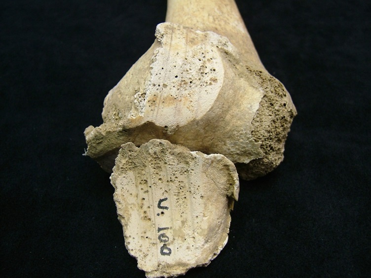
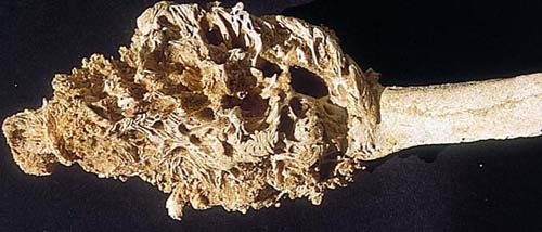
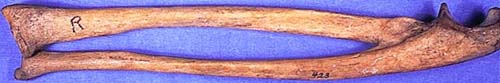
Case 144
The figure shows the inferior view of right medial clavicle, with a well-developed rhomboid fossae. Specimen found in excavation from Bouqras, Syria, ca. 8800 BP in a desert area with known exposure to the grass pea toxins.
Submitted by Dr. Deborah C. Merrett from Simon Fraser University, Burnaby, British Columbia, Canada.
Diagnosis: Case 144 Inferior view of right medial clavicle, showing a well-developed rhomboid fossae. Possible lathyrism.
Case 143
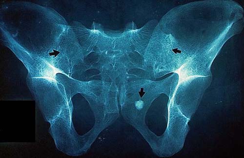
X-rays shows a lesion in a 30-year-old female mummy from Northern Chile.
Submitted by Alexander N. Gabrovsky, Virginia Commonwealth University, Medical College of Virginia, Richmond, VA.
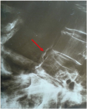
Diagnosis: Case 143 Calcification in the Abdominal Aorta.
Case 142
Skeletal lesion from a Roman period Burial. Distal femoral joint surface and patella.
Submitted by Dr. Robert R. Paine from Texas Tech University, Lubbock, TX.
Diagnosis: Case 142 Femoral joint. Degenerative joint disease.
Case 141
Skin: (Left) Right pretibial region. Young adult male, Chimu culture (Late Intermediate Period, ca. 1100-1400 AD); (Right) Microscopic picture of the skin.
Submitted by Jordi Esteban, MD and Pedro L. Fernandez, MD, PhD, from Dept. of Pathology, Hospital Clínic of Barcelona and University of Barcelona.
.jpg)
Diagnosis: Case 141 Tattoo: Black pigment in deep dermis.
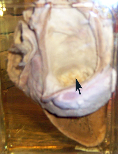
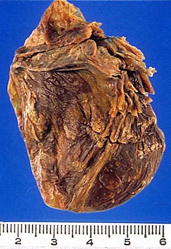
Case 140
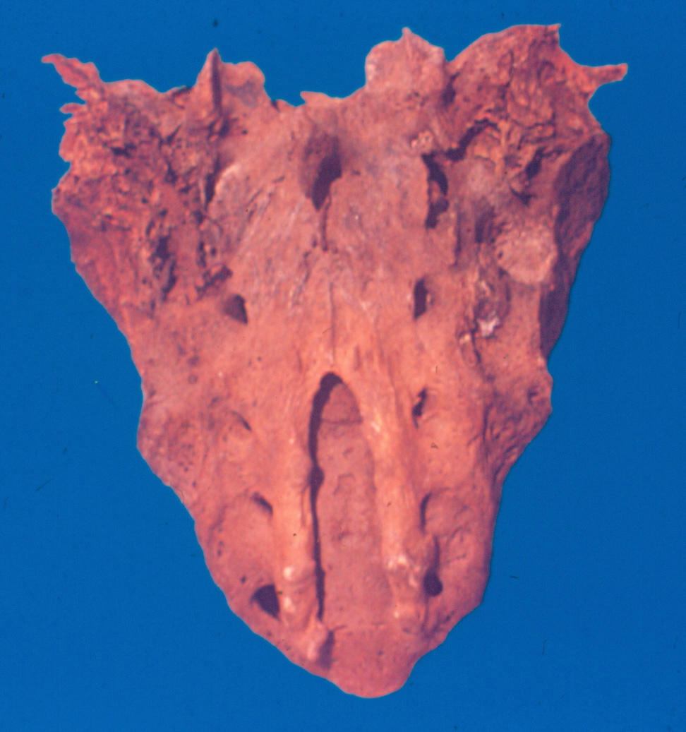
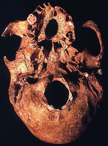
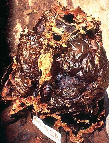
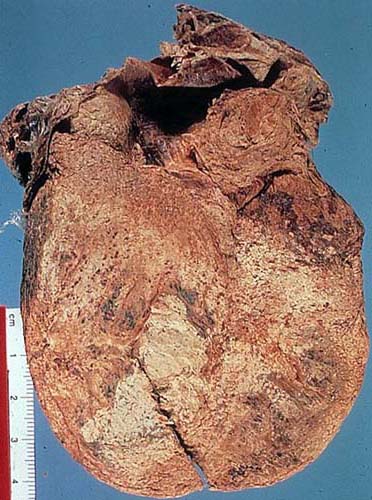
Pre-Columbian adult mummy from Northern Chile (AZ 71-T57). Organ from the thorax.
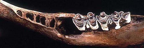
Submitted by Dr. Enrique Gerszten, Virginia Commonwealth University, Medical College of Virginia, Richmond, VA.
Diagnosis: Case 140 Heart.
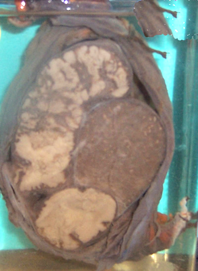
Case 139
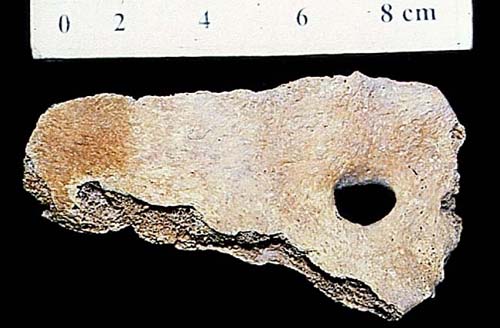
Slide shows a microscopic finding in the kidney of a South American mummy.
Submitted by Dr. Enrique Gerszten, Virginia Commonwealth University, Medical College of Virginia, Richmond, VA.
Diagnosis: Case 139 Blood vessel.
Case 138
Endoscopy of internal thoracic wall of male mummy. Location: Chiu-Chiu region (Chile). (AD VI).
Submitted by Agustín Franco, Dept. of Urology, Hospital Clinic, Barcelona; Jordi Esteban, Dept. of Pathology, Hospital Clinic, Barcelona; José Antonio Sanchez, Sc
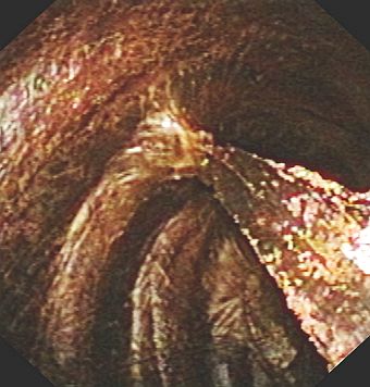
Diagnosis: Case 138 Pleural adhesions.
Case 137
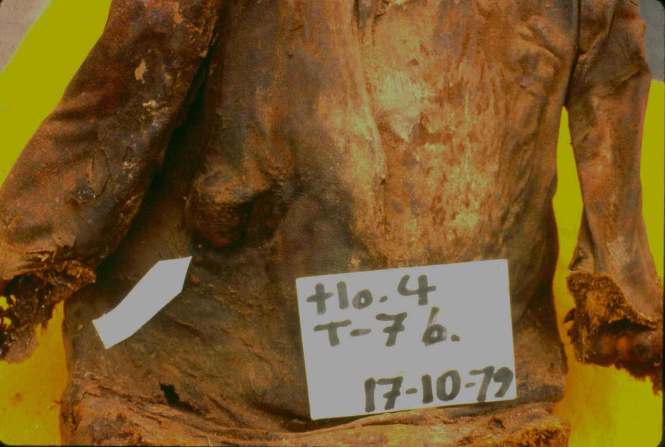
Remains of a 32-year-old woman from the Inca period (AD 1500). The lesion involves T9 through T12 with complete destruction of the vertebral bodies of T10 and T11. There is some evidence of bony fusion.
Submitted by Dr. Peter C. Gerszten, University of Pittsburgh, Pittsburgh, PA.
Diagnosis: Case 137 Healed Pott’s disease.
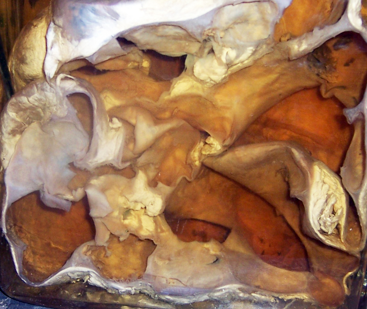
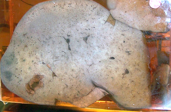
Case 136
Internal abdominal organ from a mummy from Northern Chile.
Submitted by Dr. Enrique Gerszten, Virginia Commonwealth University, Medical College of Virginia, Richmond, VA.
Diagnosis: Case 136 Spleen
Case 135
Endoscopic image from the interior of the cardiac cavity of a mummy of the region of Chiu-Chiu, Chile.
Submitted by Drs. Jodi Esteban and Pedro L. Fernández, Depto. Anatomia Patologica, University of Barcelona, Spain.
Diagnosis: Case 135 The image shows necrophagous insects belonging to the order of coleoptera (family: dermestidae).
Case 134
Skull from an excavation in Peru from the Nazca culture. The surgical technique for trephination in the skull was called crosscut and it was done following a trauma to the skull.
Submitted by Dr. Enrique Gerszten, Virginia Commonwealth University, Medical College of Virginia, Richmond, VA.
Diagnosis: Case 134 Severe periostitis following a trephination of the skull.
Case 133
The slide is from a male captive from the Pyramid of the Moon, Moche River Valley, Northern Peru, excavated from an Archeological site.
Submitted by: Dr. John Verano, Tulane University, New Orleans, LA.
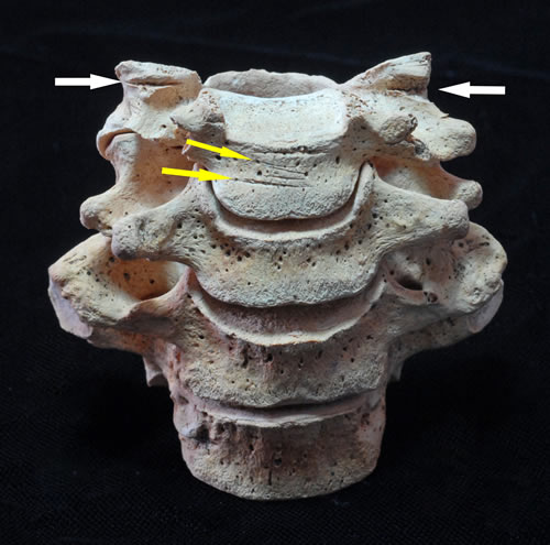
Diagnosis: Case 133 The slide shows shallow cut marks from slitting of the throat (yellow arrows) and deeper cuts, probably from decapitation of the victim.
Case 132
Lungs from a Native American from an excavation found in Southern Peru (AD 1600).
Submitted by: Dr. Enrique Gerszten, Virginia Commonwealth University, Medical College of Virginia, Richmond, VA.
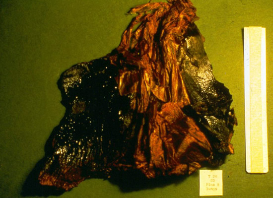
Diagnosis: Case 132 The lungs show severe anthracosis from a Native South American working in a mine.
Case 131
Skull from a 12-year-old boy from an excavation from Northern Chile (AD 1100).
Submitted by: Dr. Enrique Gerszten, Virginia Commonwealth University, Medical College of Virginia, Richmond, VA.
Diagnosis: Case 131 Biparietal porotic hyperostosis resulting from chronic anemia in a 12-year-old boy of the regional culture (AD 1100).
Case 130
Autopsy finding in a mummy of ancient Peru (young girl).
Submitted by: Dr. Enrique Gerszten, Virginia Commonwealth University, Medical College of Virginia, Richmond, VA.
Diagnosis: Case 130 Skull of a young girl with a large left parietal fracture resulting in a massive subdural hemorrhage.
Case 129
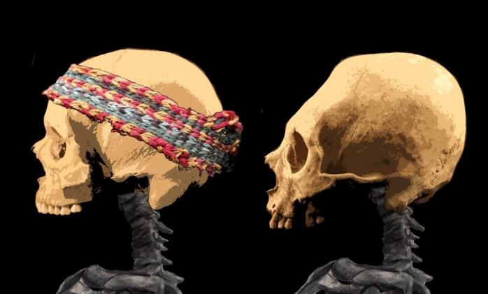
History: Pre-Columbian skull from Chile.
Submitted by: Dr. Enrique Gerszten, Virginia Commonwealth University, Medical College of Virginia Campus, Richmond, VA, USA.
Diagnosis: Case 129 Skull after artificial cranial deformation using the circular bandage technique.
Case 128
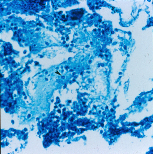
Autopsy finding in the pleura of a Pre-Columbian mummy. There were also similar nodules in the lung parenchyma.
Submitted by: Dr. Enrique Gerszten, Virginia Commonwealth University, Medical College of Virginia Campus, Richmond, VA, USA.
Diagnosis: Case 128 Miliary tuberculosis.
Case 127
Autopsy finding in the abdomen of a Pre-Columbian mummy.
Submitted by: Dr. Enrique Gerszten, Virginia Commonwealth University, Medical College of Virginia Campus, Richmond, VA, USA.
Diagnosis: Case 127 Mesenteric blood vessels.
Case 126
H&E of mummified lung from a 7 year-old boy from Castille, XIV Century.
Submitted by: Jordi Esteban, Agustín Franco*, Pedro L. Fernández. Depts. of Pathology and Urology*, Hospital Clínic, Barcelona, Spain.
Diagnosis: Case 126 Anthracosis, most likely due to fireplace smoke.
Case 125
X-Ray of a young child from a Pre-Columbian burial.
Submitted by: Dr. Enrique Gerszten, Virginia Commonwealth University, Medical College of Virginia Campus, Richmond, VA, USA.
Diagnosis: Case 125 Harris lines.
Case 124
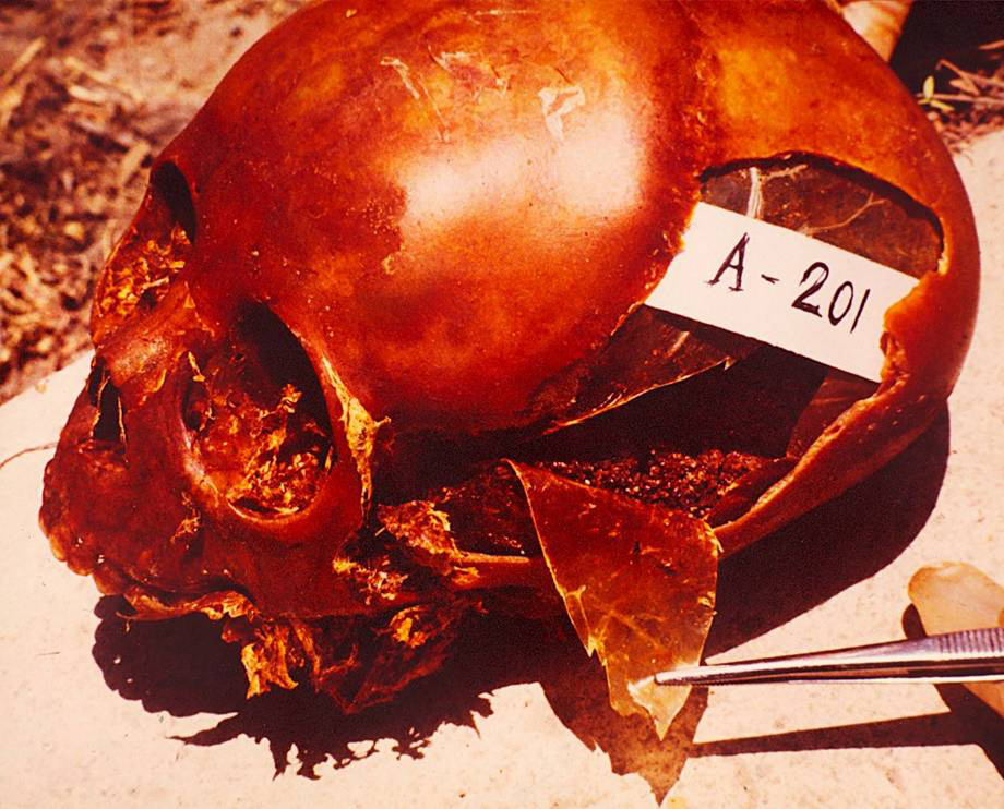
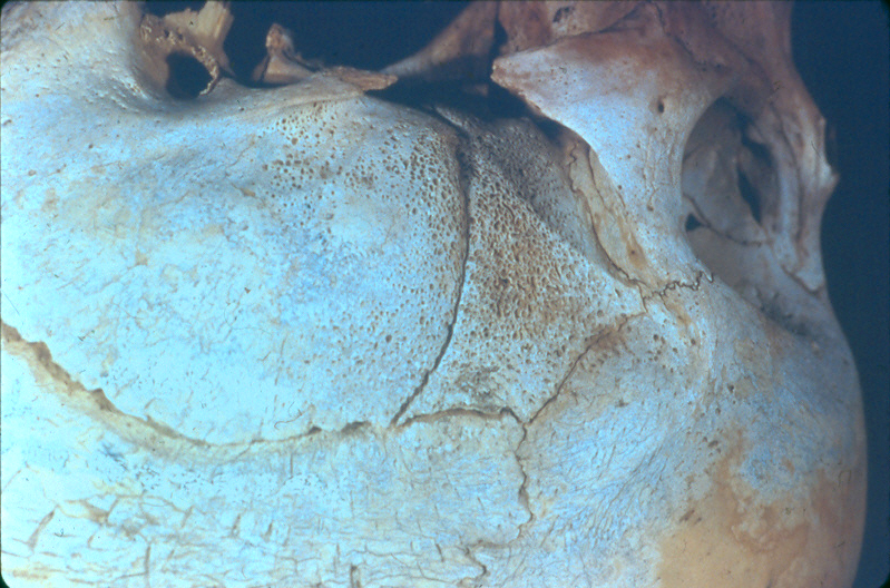
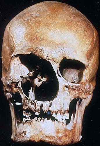
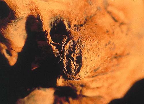
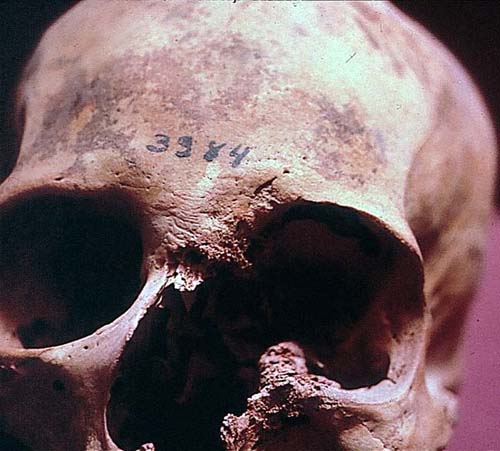
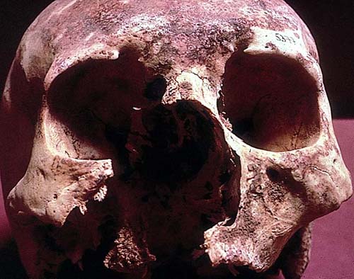
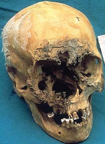
Skull from a Pre-Columbian mummy from the Azapa culture (1000 B.C.) of Northern Chile.
Submitted by: Dr. Enrique Gerszten, Virginia Commonwealth University, Medical College of Virginia Campus, Richmond, VA, USA.
Diagnosis: Case 124 Periorbital osteitis resulting from a projectile point wound to his forehead.
Case 123
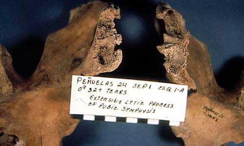
Sacrum from the remains of a 25-year-old woman from the Azapa culture of Northern Chile (1000 BC).
Submitted by: Dr. Enrique Gerszten, Virginia Commonwealth University, Medical College of Virginia Campus, Richmond, VA, USA.
Diagnosis: Case 123 Spina Bifida Occulta.
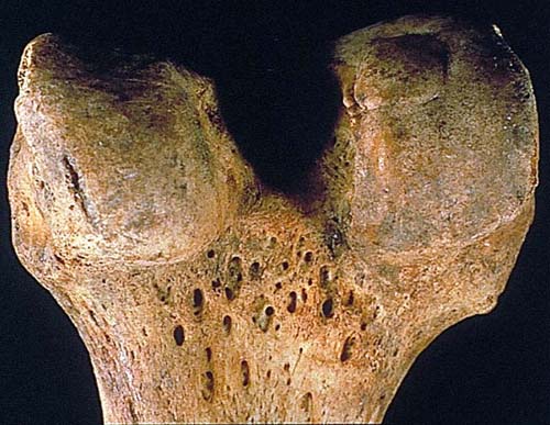
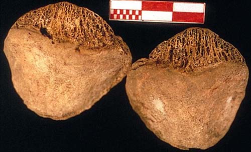
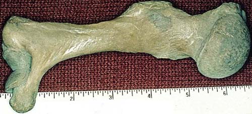
Case 122
No clinical history is available. Lesion in the head of the femur. From the Museum of Pathology.
Submitted by: Dr. Enrique Gerszten, Virginia Commonwealth University, Medical College of Virginia Campus, Richmond, VA, USA.
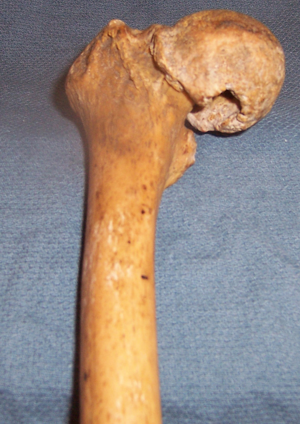
Diagnosis: Case 122 Hypertrophic osteoarthropathy affecting the head of the femur. This is a degenerative disease, marked by atrophy and degeneration of weight-bearing parts of the joint together with hyperplasia of other parts of the joint. In this case, we note atrophy and osteoporosis of the superior (articulating) surface of the head of the bone, together with the ossification of the exuberant over-growth of the cartilaginous edges around the inferior surface of the head of the femur.
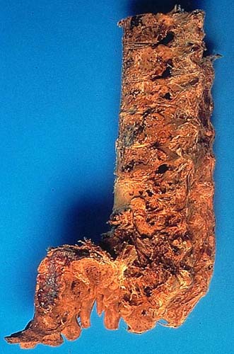
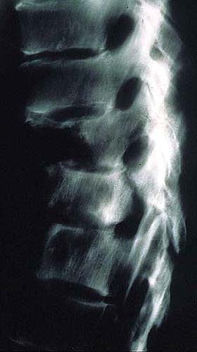
Case 121
No clinical history is available. Lateral view of a spinal column. (From the Museum of Pathology).
Submitted by: Dr. Enrique Gerszten, Virginia Commonwealth University, Medical College of Virginia Campus, Richmond, VA, USA.
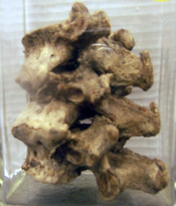
Diagnosis: Case 121 Osteo-arthritis of the vertebral column, showing thinning of the spaces between vertebrae once occupied by the intervertebral discs which have degenerated. Osteophyte formation is prominent with bony fusion apparent in some areas.
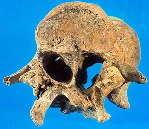
Case 120
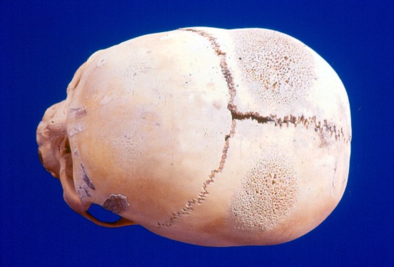
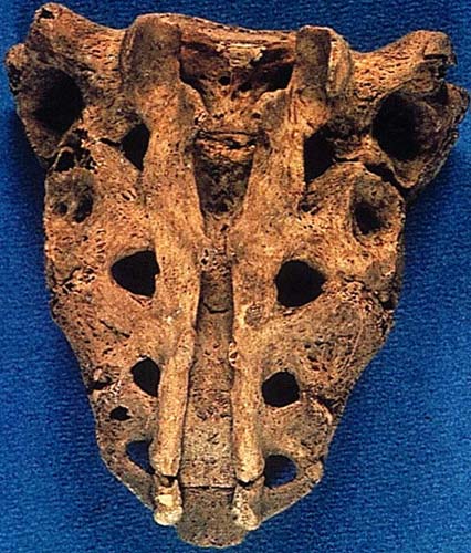
History: Skull from a 35-year-old woman of the Morro culture (6000 BC) from Northern Chile.
Submitted by: Dr. Peter C. Gerszten, University of Pittsburgh, Department of Neurosurgery, Pittsburgh, PA, USA.
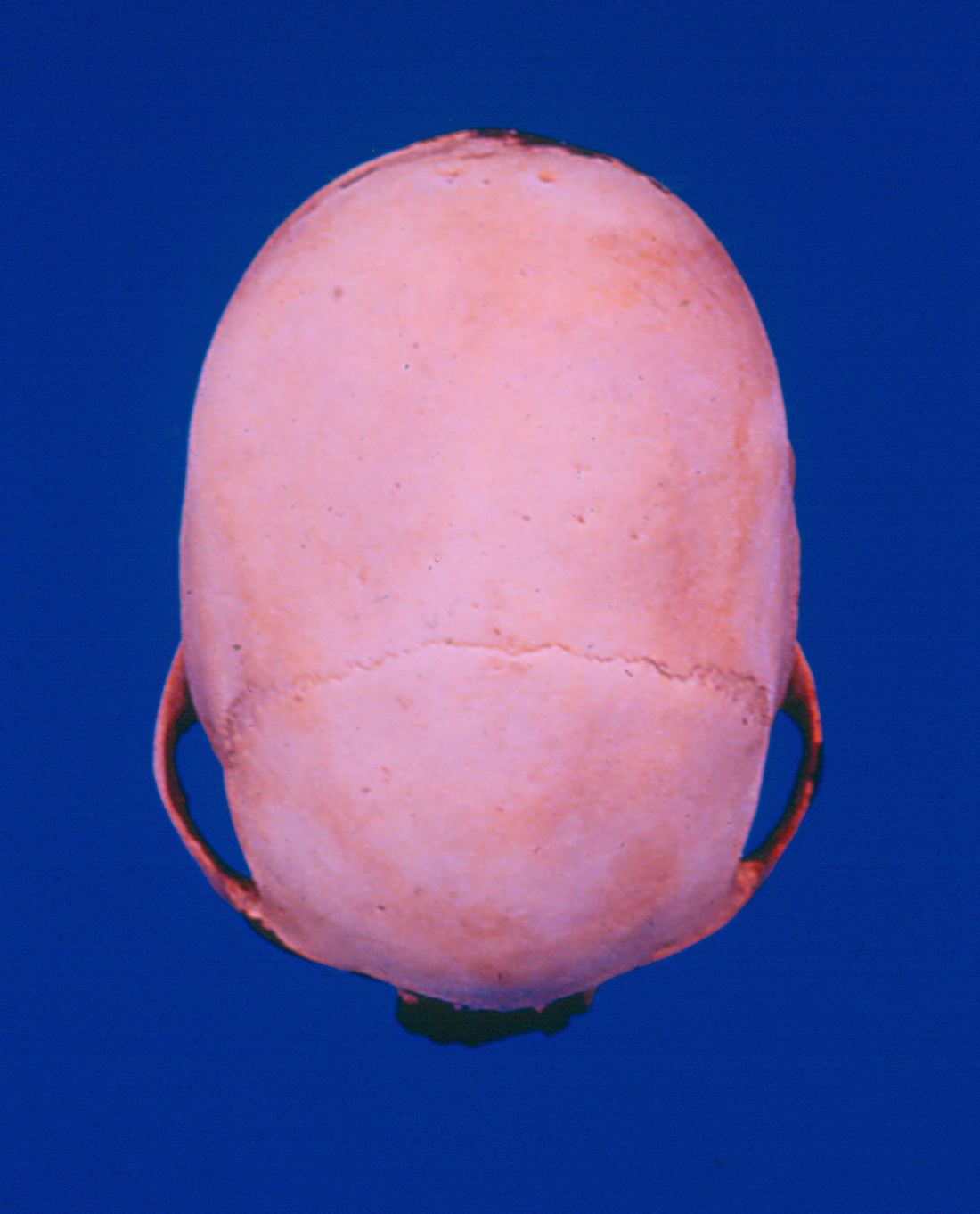
Diagnosis: Case 120 Craniosynostosis with complete absence of the sagittal sutures.
Case 119
History: Year 1920: 30-year-old female with chorea at the age of 9 with increasing cardiac failure in recent years. The left auricular wall has been cut away to show the small button-hole like orifice of the mitral valve.
Submitted by: Dr. Enrique Gerszten, Virginia Commonwealth University, Medical College of Virginia Campus, Richmond, VA, USA.
Diagnosis: Case 119 Stenosis of mitral valve due to rheumatic fever.
Case 118
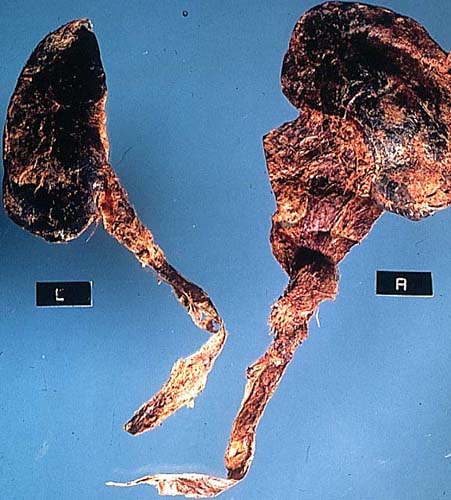
History: Year 1910: 49-year-old male with intermittent hematuria for 10 years, large mass in left flank.
Submitted by: Dr. Enrique Gerszten, Virginia Commonwealth University, Medical College of Virginia Campus, Richmond, VA, USA.
Diagnosis: Case 118 Kidney - Marked hydronephrosis.
Case 117
History: Year 1900: 55-year-old male with a long history of chronic disease.
Submitted by: Dr. Enrique Gerszten, Virginia Commonwealth University, Medical College of Virginia Campus, Richmond, VA, USA.
Diagnosis: Case 117 Tuberculosis of the epididymis.
Case 116
History: 366639 National Museum of Natural History, Smithsonian Institution. Skull with slight postmortem damage. Postcranial elements include both clavicles and the right scapula all of which are normal. The burial is from the Uyak Site on Kodiak Island, Alaska. It was accessioned by Dr. Aleš Hrdlicka in 1932. Hrdlicka thought the burial was from the middle pre-Koniag period which, if correct, would have a date well before historic Russian contact. The skull is from a female around thirty years of age at the time of her death.
Submitted by: Dr. Donald Ortner, National Museum of Natural History, Smithsonian Institution, Washington, DC.
Diagnosis: Case 116 Hansen’s disease (Leprosy).
Because of the combination of remodeling of the margins of the piriform aperture with destructive remodeling of the anterior hard palate, the most likely diagnosis is leprosy. Treponematosis and tuberculosis can cause similar lesions but remodeling of the anterior hard palate would be unusual.
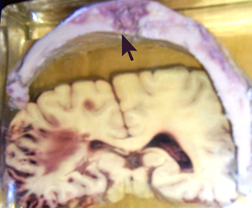
Case 115
History: A 46-year-old white, obese female with frontal headaches for a month. She had a sudden onset of dizziness and fainting, which caused her to fall down some stairs. She was unconsciousness until death, (A2892-1939).
Submitted by: Dr. Enrique Gerszten, Virginia Commonwealth University, Medical College of Virginia Campus, Richmond, VA, USA.
Diagnosis: Case 115 Large Cerebral aneurysm.
Large aneurysm (giant aneurysm) of the right middle cerebral artery, pressing into the right cerebral hemisphere. The aneurysm has been cut across to show mass of blood clot. There was rupture of the aneurysm and massive subarachnoid hemorrhage.
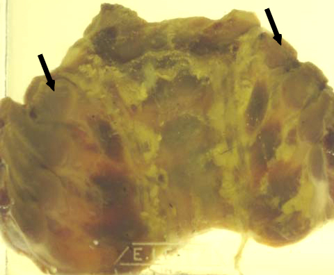
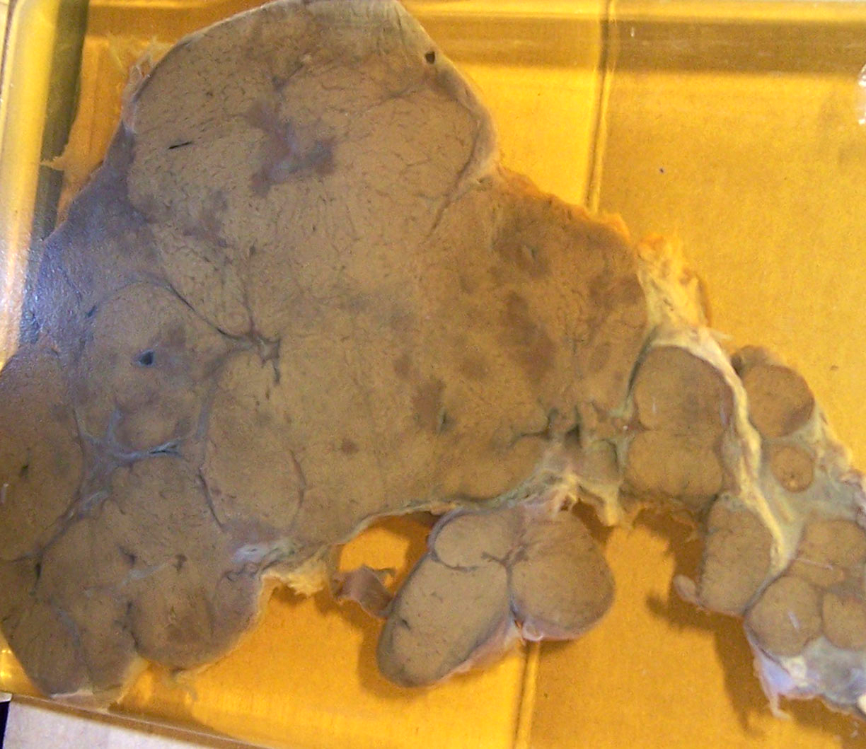
Case 114
History: From a white male age 46, who had noticed an enlarging abdomen for 8 years, but was otherwise well (died in 1930).
Autopsy finding: Picture showing an immense spleen weighing 5,890 gms. and measuring 38x20x13 cms., which completely filled the entire left side of the abdominal cavity. Its surface was smooth, deep blue in color, with white spots irregularly scattered over it.
On section the splenic substance was dark red and firm. The splenic vessels were much enlarged and tortuous.
Submitted by: Dr. Enrique Gerszten, Virginia Commonwealth University, Medical College of Virginia Campus, Richmond, VA, USA
Diagnosis: Case 114 Gaucher disease.
Microscopically the entire spleen was occupied by Gaucher cells with large round, pale cytoplasm and small darkly stained round nucleus excentrically placed. This case was published in the Am. J. of Medicine and Science, 1935, Vol. 190, pg. 511.
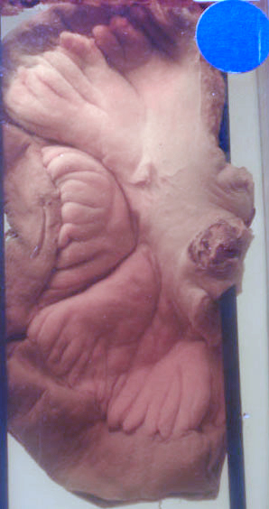
Case 113
History: Incidental finding in an autopsy of a 65-year-old male who died in an accident (1920).
Submitted by: Dr. Enrique Gerszten, Virginia Commonwealth University, Medical College of Virginia Campus, Richmond, VA, USA
Diagnosis: Case 113 Paget Disease.
Case 112
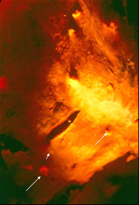
History: This section is from the liver of a female mummy. She died three thousand years ago in the Northern Chilean valley of Tarapaca. Her liver was one of 50 used to detect virus diseases using monoclonal antibodies and DNA probes in a detection system of DAKO.
Submitted by: Dr. Marvin J. Allison, Virginia Commonwealth University, Medical College of Virginia Campus, Richmond, VA, USA
Diagnosis: Case 112 Cells containing Epstein-Barr Virus
Case 111
History: This hand is from an autopsy from a Virginia farmer, around 1890, from the Pathology Museum of our Department. These lesions are from areas of the body exposed to the sun. His clinical history also revealed diarrhea and dementia.
Submitted by: Dr. Enrique Gerszten, Virginia Commonwealth University, Medical College of Virginia Campus, Richmond, VA, USA
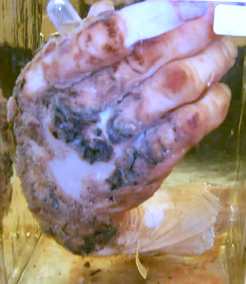
Diagnosis: Case 111 Pellagra (niacin or a tryptophan deficiency).
Case 110
History: Hematoxilin and eosin staining of an internal organ of a 40-yer-old male mummy from the Azapa Valley (1,000 A.D.). Dx: Identify the tissue?
Submitted by: Pedro L. Fernández, Jordi Esteban and Montserrat Tortosa, Dept. of Pathology, Hospital Clínic, University of Barcelona, Spain.
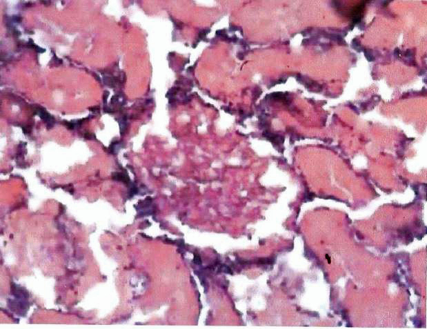
Diagnosis: Case 110 Kidney.
Case 109
History: Sixty out of 92 skeletons from Pre-Columbian natives from the Costal San Diego area shows this common dental abnormality.
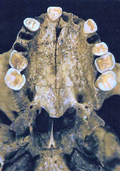
Diagnosis: Case 109 Dental attrition. Submitted by: Tori Heflin, University of Cambridge, Cambridge, UK
Case 108
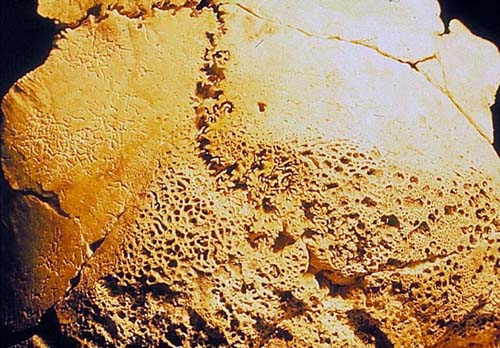
The picture shows a segment of the internal surface of the frontal bone with a slightly thickened diploé and a marked bone rearrangement of the inner table at the level of the frontal crest. At both sides of the great falx cerebri, starting from the orbitary vault until the groove of the median meningeal artery, symmetric thickenings have developed in form of plaques or festoons. In the central portion, at the level of the metopic suture, an area of eburnation, also symmetrical, can be observed.
Osteological Collection from the Museum of Anthropology and Ethnography of Turin (Italy). This specimen comes from S. Samuele Church (Venezia, Italy) XVII-XVIII Century P.C. Sex: female. Age: adult.
Pictures showing internal view of a frontal bone with osseous reaction in the frontal crest
Submitted by: Dr. Francesco Merlo, Dr. Rosa Boano, Prof. Ezio Fulcheri
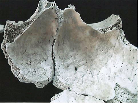
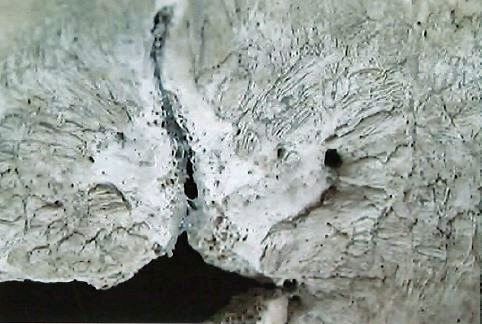
Diagnosis: Case 108 Hyperostosis frontalis interna, also known as Morgagni-Stewart-Morel Syndrome.
Case 107
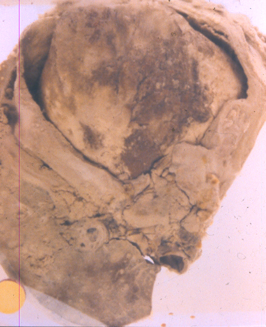
Posterior view of a skull with cranial deformation from Pre-Columbian times recovered from a cemetery in Paracas, Peru. What is your diagnosis?
Submitted by: Garry Kerr, University of Montana, Missoula, Montana, USA.
Diagnosis: Case 107 Chronic infection of unknown etiology.
Case 106
Histopathology of a liver from a 10-year-old girl who died 1800 years ago in the coastal area that is now near Arica, Chile. The slide is an ELISA with a specific antibody using FAST RED of an infectious disease that was prevalent in the same area until 60 years ago. What is your tentative diagnosis?
Submitted by: Dr. Marvin J. Allison, Virginia Commonwealth University, Medical Campus, Richmond, Virginia, USA.
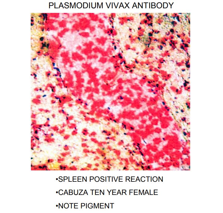
Diagnosis: Case 106 Malaria.
Case 105
Internal aspect of anterior thorax of a child (year 1900, Virginia, USA).
Submitted by: Dr. Enrique Gerszten, Virginia Commonwealth University, Medical Campus, Richmond, Virginia, USA.
Diagnosis: Case 105 Rickets (Rachitic rosary).
Case 104
Autopsy finding in the liver, late stage of an infection (year 1900, Virginia, USA).
Submitted by: Dr. Enrique Gerszten, Virginia Commonwealth University, Medical Campus, Richmond, Virginia, USA.
Diagnosis: Case 104 Tertiary syphilis of the liver (Hepar lobatum).
Case 103
Incidental finding in an autopsy performed in the year 1900 in Virginia. Nodule in the mesentery.
Submitted by: Dr. Enrique Gerszten, Virginia Commonwealth University, Medical Campus, Richmond, Virginia, USA.
Diagnosis: Case 103 Tuberculosis, calcified mesenteric lymph node. This is frequently seen after bovine tuberculosis, which has disappeared in the USA for two generations due to tuberculin testing in cattle and extermination of infected animals.
Case 102
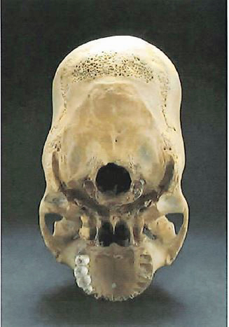
This skull is from a male about 35 years old. The specimen is from a coastal Peruvian population that lived 1000 years ago with a marine economy. This cemetery was hit by a number of Tsunamis in the past, only human skeletons were found.
Submitted by: Dr. Marvin J. Allison, Virginia Commonwealth University, Medical Campus, Richmond, Virginia, USA.
Diagnosis: Case 102 The ossified styloid process is elongated above normal level and may possibly produce the Eagle syndrome.
Case 101
Slide showing a skull from an adult male. There is a defect in the right frontal bone. The defect is about 1 cm. The skull was excavated in Ermita de Valgañón, Spain (XIII Century).
Submitted by: Dr. J. Martínez Flórez, Departamento de Antropología, Museo de La Rioja, Logroño (La Rioja, España).
.jpg)
Diagnosis: Case 101 Trephination on right frontal bone.
Case 100
Slide showing lateral view of maxilla with an osseous lesion on the alveolar process. Etruscan-Roman Craniological Collection (VIII-I century B.C.) from the Museum of Anthropology and Ethnography of Turin (Italy). Archaeological specimens excavated from the Tarquinia necropolis (Viterbo, Lazio, Italy). Sex of specimen: Male. Age at death: adult, 40-45, years old.
Submitted by: Dr. Rosa Boano, Dr. Patrizia Rizzo, Prof. Emma Rabino Massa, Prof. Ezio Fulcheri
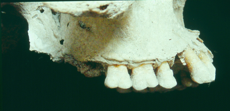
Diagnosis: Case 100 Inflammatory chronic process on external and internal surface of maxilla with osseous reaction: alveolar pyorrhea.
Case 99
From a white male aged 16, with pain in the left chest anteriorly, headache, malaise, a non-productive cough. Three weeks later developed night sweats, dyspnea, precordial pain, with a weight loss of 30 lbs.; temperature102; dullness in the left base posteriorly with diminished breath sounds. Sputum showed Gram-positive filaments. X-ray reported atypical pneumonia extending out from the root of the left lung practically to the periphery. (From the Pathology Museum). Died in 1944.
Submitted by: Dr. Enrique Gerszten, Medical College of Virginia Campus, Richmond, Virginia, USA.
Diagnosis: Case 99 Actinomycosis.
Case 98
This 1000-year-old ceramic found in Peru shows an activity that is still done in exactly the same manner in rural Peru and Bolivia. What are they doing?
Submitted by: Dr. Marvin J. Allison, Medical College of Virginia Campus, Richmond, Virginia, USA.
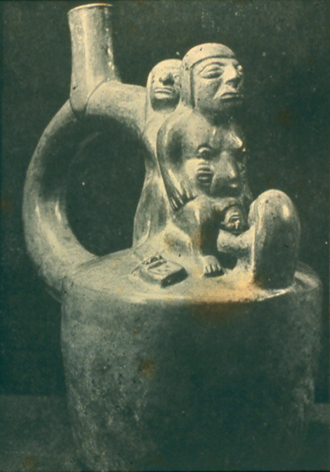
Diagnosis: Case 98 The ceramic is about 1,000 years old and shows a woman delivering a baby. She is standing supported by presumably her relatives and the midwife is squatting in a hole in the ground to catch the baby as it comes out. If the baby is slow to deliver, often a 3” broad sash is tied above the swollen abdomen and tightened with a stick. Archeologists have found several female mummies with the sash still in place and the woman died of hemorrhage. This system is still in use in rural areas of the Andes today.
Case 97
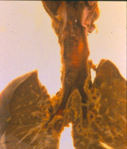
This is a section of the intestine from a Chinese contract laborer in 1853 (Case #96 was a section from his liver). He arrived sick at the Peruvian immigrant quarantine station on San Lorenzo Island. He was hospitalized there and died before he could enter continental Peru.
Submitted by: Dr. Marvin J. Allison, Medical College of Virginia Campus, Richmond, Virginia, USA
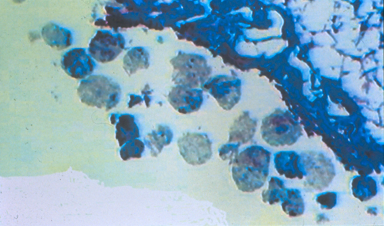
Diagnosis: Case 97 This Chinese contract laborer who died in Peru in 1853 had 3 parasites, Schistosoma japonicum in the liver, Paragonimus in the lung, and Balantidium coli in the colon. The immediate cause of death was due to an acute enteritis resulting from the B. coli infection. This pig parasite is the largest protozoa infecting man and is common in children in rural areas of modern Peru. We assume he picked this parasite up while in the Peruvian hospital on the island of San Lorenzo and his two chronic diseases had already weakened him severely.
Case 96
This is a section of the liver from a Chinese coolie contract laborer in 1853. He arrived sick at the Peruvian immigrant quarantine station on San Lorenzo Island. He was hospitalized there and died before he could enter continental Peru.
Submitted by: Dr. Marvin J. Allison, Medical College of Virginia Campus, Richmond, Virginia, USA.
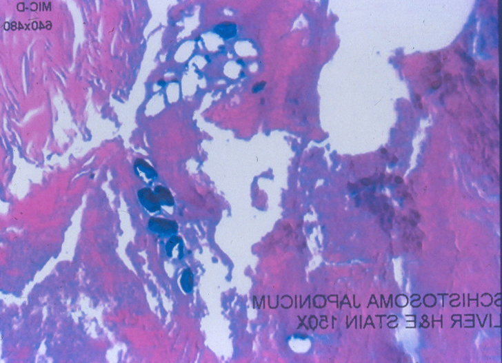
Diagnosis: Case 96 Schistosoma Japonicum infection showing several nests of eggs in the liver.
Case 95
This is a modern historical case, around 1900, from the Pathology Museum of our institution. His clinical history was that of a black, male child with nausea, vomiting, lassitude, fever, cough, and difficulty in breathing for one week before admission to the hospital. His respiration became more labored with intercostal retraction, pulse 125, respiration 36, temperature 100.4 with pallor. A tracheotomy was done with some improvement, but the tube became plugged on several occasions. After 4 days he developed convulsions, cyanosis and died.
Submitted by: Dr. Marvin J. Allison, Medical College of Virginia Campus, Richmond, Virginia, USA.
Diagnosis: Case 95 Diphtheria.
This was a case of diphtheria from our Departmental Museum around 1900 and should be considered one of the historic diseases nearly conquered by DPT vaccine administered to children. Strangely enough there has been a reactivation of this disease in Germany in recent years and it is still found in developing areas around the world.
Case 94
Slide showing typical lesions seen on the skin of a leg from a mummy of the Valley of Camarones (Northern Chile) about 500 before present. In the same archeological excavation numerous mummies with the same type of lesions were seen on the limbs and the thorax.
Submitted by: Dr. Marvin J. Allison, Medical College of Virginia Campus, Richmond, Virginia, USA.
Diagnosis: Case 94 Chronic Arsenic Dermatitis.
This case is one of the 27 mummies with chronic arsenic poisoning from drinking the water in the valley of Camarones, Chile. These lesions are hypo and hyperpigmentation and are associated with 2-3 grams of arsenic accumulated in the skin. The arsenic concentration in the body depends upon the daily dose and in a Mexican study lesions such as these took 8-10 years to appear on the skin, in 25 years and with 8 grams of arsenic in the skin you would see papular keratosis, and after 38 years and over 10 grams of arsenic appears ulcerative lesions and skin carcinomas. Only one of the 27 cases had a skin carcinoma. A study on modern inhabitants of the valley showed they drank daily 2.5 mgms. of arsenic and 80% of the modern inhabitants had lesions like their pre-Columbian forefathers with also one case of skin cancer. There were numerous fresh water springs, almost free of arsenic, but the modern people preferred to drink the river water stating that it tastes better.
Case 93
This slide is from a 6-year-old boy who died in Ilo, Peru area, about 1,200 years ago.
A. Radiograph of liver
B. Calcium stain of liver section (100X)
C. H&E stain of G.I. tract of parasite
Submitted by: Dr. Marvin J. Allison, Medical College of Virginia Campus, Richmond, Virginia, USA.
Diagnosis: Case 93 Ascaris lumbricoides.
Case 92
This case is that of a 23-25 year old woman who died in childbirth 1,000 years ago in southern Peru. She had a large blood clot the size of a baseball in her perineal area. The symphysis pubis and sacral-iliac articulations were open. Her breasts were full with milk with a large tumor mass on the right and several small nodules on the left. An x-ray of the thoracic cavity, oblique and lateral, showed calcification in the tracheal-bronchial lymph nodes.
Submitted by: Dr. Marvin J. Allison, Medical College of Virginia Campus, Richmond, Virginia, USA.
Diagnosis: Case 92 Coccidioidomycosis.
Case 91
Fifth metatarsal bone from a medieval cemetery from the Church of Sant Joan de Benlliure, Catalonia, Spain.
Submitted by: Dr. Domingo Campillo, Museum of Archeology, Barcelona, Spain.
Diagnosis: Case 91 Diaphyseal fracture of the fifth metatarsal. The fracture is also known as Jones fracture.
Case 90
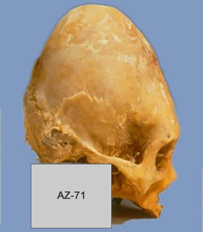
Top view from a skull of a 21-year-old female from the Gentilar culture (AZ-8, T 38) from 1200 to 1400 AD found in the Azapa Valley, Arica, Chile.
Submitted by: Dr. Peter C. Gerszten, Department of Neurosurgery, University of Pittsburgh, Pittsburgh, PA 15213. USA.
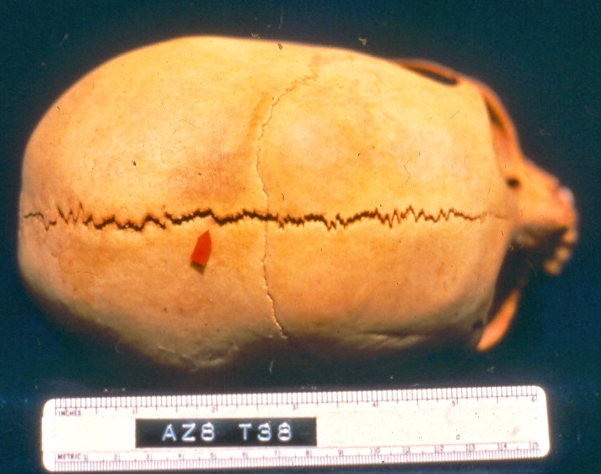
Diagnosis: Case 90 Metopic and Sagittal sutures open due to intentional cranial deformation.
Case 89
Left lateral skull with abnormal porosity of the greater wing of the sphenoid and temporal bone in a child about eleven years of age. The skull is from an archeological site in Pachacamac, Peru, dated between 900 and 1450 AD. The lesion is bilateral and also is associated with bilateral lesion on the orbital roof and the posterior maxilla and zygomatic bone.
Submitted by: Dr. Donald J. Ortner, Department of Anthropology, Smithsonian Institution, Washington, DC 20560-0112, USA
Diagnosis: Case 89 Scurvy.
Case 88
A six year old girl who died in 800 AD from Southern Peru (Hilo).
Submitted by: Marvin J. Allison, Medical College of Virginia Campus, Richmond, Virginia, USA.
A – Close up of lower face showing the nasal area exposed by a disease process.
B – Microscopic area of the lesion of skin of the adjacent nasal area (Giemsa stain)
Diagnosis: Case 88 Mucocutaneous lesion of uta. Probably due to leishmaniasis peruensis/braziliensis.
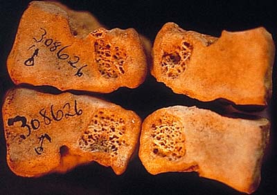
Case 87
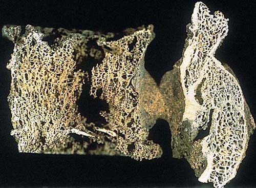
Transversal midsections of femoral bones of Modern and Stone Age (5,000 BP) women.
Submitted by: Dr. Pia Bennike, Laboratory of Biological Anthropology, University of Copenhagen, Denmark.
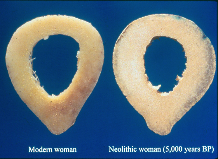
Diagnosis: Case 87 This slide is of two transversal midsections of femoral bones. They are showing an average difference of bone area (compact cortical thickness) and marrow cavity of modern Danish women (left) and Neolithic women (5,000 years BP). It should be noticed that the section of the Neolithic femur (right) has a slightly smaller marrow cavity and a larger compact cortical thickness.
Case 86
Skin from a mummy excavated in the Camarones Valley from the Inca culture in Northern Chile. Slide shows a scanning microscopic picture and a section stained with H&E of a protruding lesion.
Submitted by: Jordi Esteban, Pedro L. Fernandez, Montserrat Tortosa, Elisenda Coll+, Josep Palout# from the Dept. of Anatomical Pathology and #Dermatology, Hospital Clinic, IDIBAPS, +Microscopy Unit, SCT, University of Barcelona, Spain.
Diagnosis: Case 86 Foliculitis.
Case 85
This image shows an inferior view of a maxilla (palatine and alveolar process) from a fragmented and incomplete skeleton.
Submitted by Fulcher, E., Boano, R., and Panto, G.
Diagnosis: Case 85 Ectopic tooth.
Case 84
This image shows an H&E section from an eight-year-old girl. She was excavated from a Huari Gallery Burial in rural Nazca, Peru. She died about 1,200 years ago and was accompanied in death by 11 adults buried in a replica of a mountain house.
Among the dead in the house were thrown a beheaded man, woman and two children to serve the ceremonial dead. These were probably taken as prisoners since they were not ceremonial burials. The mummies were wrapped with many pounds of cotton and an outer cloth. The bundles had a false head, and some had masks.
The young girl's liver was one of 40 used in a viral hepatitis study in pre-Columbian mummies. The H&E section shows numerous tunnels in the liver. The material in the image was found in several tunnels.
Diagnosis: Case 84 Necrotic liver fluke.
Case 83
Adult female from 4th century Spain with alteration of the posterior surface of L3 and L4 vertebrae.
Submitted by Dr. Campillo
Diagnosis: Case 83 - Disc herniation with protrusion into the spinal canal.
Case 82
X-ray from a 30-year-old female who died 350 years ago.
Diagnosis: Case 82 Recently delivered a child.
Case 81
The gross (above) and X-ray (below) are from the Pueblo culture before the European invasion (1300-1450). They represent an infant's thoracic skeleton several weeks of age, found in place in a grave dug in the floor of a room in a Pueblo dwelling.
Submitted by Dr. C. F. Merbs
Diagnosis: Case 81 Birth trauma.
Case 80
The image shows the sternum of a female 35-39-years-old who died 800 BP in the Columbian Andes.
Submitted by Dr. Felipe Cardenas-Arroyo
Diagnosis: Case 80 Congenital anomaly of the sternum.
Case 79
Skull of undetermined sex--14th-15th century. The skull presents a post-mortem fracture that exposes the ethmoidal sinus in which was seen a little pedunculated lesion, 0.8 cm in its greatest diameter.
Submitted by E. Fulcheri, L. Ferrari, S. Boccone & R. Boano
Diagnosis: Case 79 Chronic rhinosinusitis.
Case 78
Adult prehistoric female from Chilca, Peru. Image shows the sacro-iliac region.
Submitted by Rose Tyson
Diagnosis: Case 78 Tuberculosis.
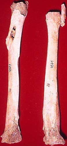
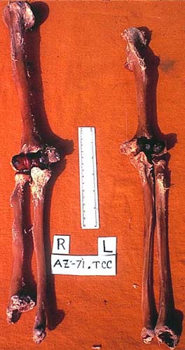
Case 77
Adult female from pre-historic Lomas, Peru. Image shows right tibia and fibula (anterior), and right femur (posterior).
Submitted by Rose Tyson
Diagnosis: Case 77 Osteomyelitis of femur, tibia and fibula.
Case 76
Section of the rectum from a mummy of a middle-aged male dating to ca. 300 AD from Dakhleh Oasis in Middle Egypt.
Submitted by Dr. Michael R. Zimmerman

Diagnosis: Case 76 Schistosomiasis.
Case 75
Biopsy of the left arm ulcer from Maria d' Aragona, Marquise of Vasto (1503-1568).
Submitted by Dr. Gino Fornaciari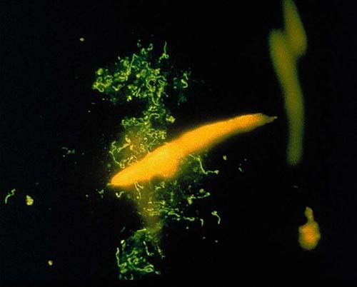
Diagnosis: Case 75 Syphilis.
Case 74
Ferrante I of Aragon, King of Naples died in 1494 at the age of 63. The autopsy evidenced in the small pelvis a fragment of hollow fibrous tissue, probably the rectum.
Submitted by Dr. Gino Fornaciari

Diagnosis: Case 74 Tumor of rectum.
Case 73
The specimen is from an adult male. His grave dates back to the 14th-15th centuries. The right femur presents, at the distal epiphysis, a well defined incision partially delimiting a depressed area.
Submitted by Dr. E. Fulcheri and Dr. R. Boano
Diagnosis: Case 73 Osteochondritis Dissecans in the necrotic phase.
Case 72
Fecal sample from a 34-year-old female who died 2,500 years ago in Northern Chile. Test with fluorescent monoclonal antibodies.
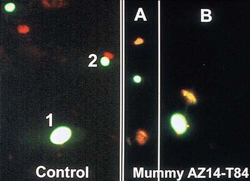
Diagnosis: Case 72 Cryptosporidium parvum (A) and Giardia lamblia (B).
Case 71
Specimen from the spine of a 20-year-old individual recovered during excavation at the site of a burial tomb in the Negev Desert of Israel. The tomb dates from the Roman period circa AD 100.
Submitted by Dr. Baruch Arensburg
Diagnosis: Case 71 Diastematomyelia of the Spinal Cord.
References: Edelson, J. G., Nathan, H., & Arensberg, B. (1987). Diastematomyelia: The double-barreled spine. Journal of Bone and Joint Surgery, 69, 8, p. 188.
Case 70
Both patellas belong to a 40-year-old potter from the Huaca de la Luna site at the Moche Valley (Peru). The remains were recovered from an urban architectonic complex of Moche IV Fase (350-600 AD).
Submitted by Mario Millones
Diagnosis: Case 70 Lesion on the internal distal face of both patella due to a kneeling position associated with pottery making.
Case 69
The image is from a breast section stained with fast red against antibody to MAM6. It is from the mummy of an 18-year-old girl belonging to the Maitas culture who died about 1000 years ago. She was our youngest pregnant female and had delivered one twin, but bled to death before the delivery of the second baby. She held the umbilical cord of the first baby in her hand.
Diagnosis: Case 69 Reactive human milk fat globule protein antigen (1000 years old).
Case 68
Male 60-65-years-old from 12-13th century in Italy. Specimen is the elbow area of the left arm.
Submitted by E. Fulcheri, M. Reto, and R. Boano
Diagnosis: Case 68 Fractures of the proximal metaphyseal part of the left radius and ulna together with fracture of the elbow joint.
Case 67
Vertebral column from a 20-year-old Peruvian man who died 1150 AD. Examination of the lumbar spine and sacrum revealed a severe retolisthesis of the lumbosacral junction with severe compression fractures of the L4 and L5 vertebral bodies. In addition, there was a loss of the normal lumbar lordosis. Radiographic examination revealed a complete bony fusion of the L3-S1 vertebral bodies. This was confirmed by computed tomography.
Submitted by Dr. Peter Gerszten
Diagnosis: Case 67 Traumatic injury.
Case 66
A 50-year-old man from the Nazca culture (southern Peru) from ± 900 AD (Lineal Tomography).
Submitted by Dr. Guido P. Lombardi
Diagnosis: Case 66 Metastatic tumor involving T10-12. Osteolytic lesions, probably from a metastatic tumor, are shown involving the vertebral bodies of the T10-T12 segment.
Case 65
Gross (above) and X-ray (below) from the heart of a 3-year-old female from the Cabuza culture (350 AD).The material injected was Hypaque 60 (meglumine diatrizoate 60%).
Submitted by Dr. Enrique Gerszten, Dr. Marvin J. Allison and Dr. James Messmer
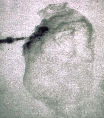
Diagnosis: Case 65 Normal coronary arteries.
Case 64
Metatarsal bone of bovine, found in a IV Century Roman area in Varea (Logroño, La Rioja) of northern Spain.
Submitted by Dr. Julio Martinez Flores
Diagnosis: Case 64 Tuberculosis of bovine metatarsal bone.
Case 63
The X-ray is from a 16-year-old female, Cabuza culture, Northern Chile (350-500 AD).
Diagnosis: Case 63 Severe chronic osteomyelitis with a large sequestrum of the humerus.
Case 62
The archeological specimen is from a young man 23-25 years of age, dated 200 BP. The lesion is on the seventh rib, right, size 11x8x7 cm. It shows a growth of bone forming a "sun burst effect" with areas of bone destruction.
Submitted by Milagros Macías López
Diagnosis: Case 62 Osteogenic sarcoma.
Case 61
EM image from the chest organ of a northern Chilean mummy. The mummy was a 12-year-old female from the Cabuza culture (300-600 AD).
Diagnosis: Case 61 EM showing bands of the myocardium in normal myocardium.
Case 60
Vertebrae found in the archeological site Kon-Kon, Rio Chillón, Lima, Perú (1100-1400 AD). The image shows two dorsal vertebrae with synostosis. In the left lateral side of the body, there is a lesion without alteration of the vertebral architecture.
Submitted by Dr. Carlos E. Ponce García
Diagnosis: Case 60 - Vertebral abscess.
Case 59
Hyoid bone with 5 mm round lesion on external surface, with smooth borders and slight bony build-up around a rim of lesion and thin bony floor at base of lesion. This bone comes from a young adult caucasian male, 20-21 years of age, from a 13th century Frankish cemetery in Corinth Greece.
Submitted by Ethne Barnes, PhD
Diagnosis: Case 59 - Thyroglossal cyst.
Reference: Moore, K. L. (1985). Clinical oriented anatomy. (2nd ed.). Baltimore: Williams & Wilkens, pp. 1026, 1029, 1066.
Case 58
An egg found in a latrine in the English settlement at Ferryland, near Saint Johns, Newfoundland, Canada. This settlement was founded by George Calvert, 1st Lord of Baltimore, in 1621.
Submitted by Patrick Horne
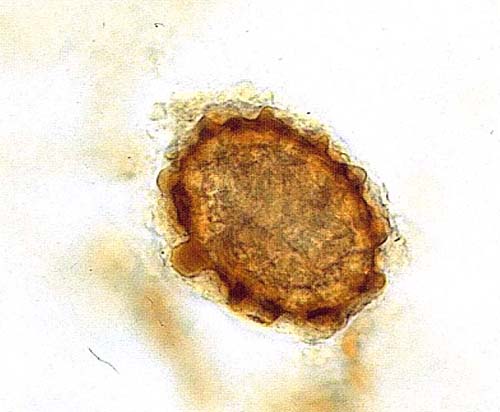
Diagnosis: Case 58 - Ascaris egg.
Case 57
This material was excavated at the Carrier Mills site in southern Illinois (dated 7000-5000 BP). The material consists of a portion of dorsal lumbar spine composed of five vertebrae and associated intervertebral disc. On cut surface, the intervertebral discs appear preserved and calcified.
Submitted by Dr. Kenneth P. H. Pritzker and Dr. Bruce Rothschild
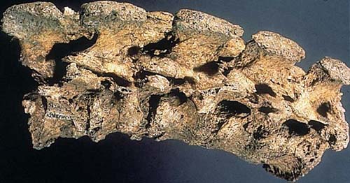
Diagnosis: Case 57 - Ankylosing spondylitis with disc calcifications.
Case 56
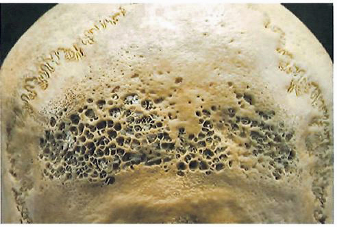
The posterior cranial vault region of an adult male Mayan from northern Belize, dating from the Postclassical period (circa 1300 AD).
Submitted by Dr. Christine D. White
Diagnosis: Case 56 - Active Porotic Hyperostosis caused by ongoing iron deficiency anemia.
Case 55
Skull of a young male from Neolithic Denmark (7000 BP).
Submitted by Dr. S. Ry Andersen
Diagnosis: Case 55 - Benign Tumor of Nasal Cavity.
Reference: Brothwell, D., and Sandison, A. T. (1967). Diseases in Antiquity. Springfield, IL: Charles C. Thomas, p. 329.
Case 54
Specimen from the Monterey Bay area of Central California dates from 2200-4900 BP. The lesion is a rough, oval-shaped defect involving the lateral cortex of the mastoid process of the left temporal bone, extending from the mastoid tip to just cephalad to the floor of the external auditory canal.
Submitted by Lorna C. Pierce, PhD
Diagnosis: Case 54 - Acute otitis media complicated by mastoiditis.
Reference: Loveland, C. J., Pierce, L. C., Gregg, J. B. Ancient temporal bone osteopathology, San Jose State University, San Jose, CA, pp. 95192-0113
Case 53
The picture shows the skull of a 4-year-old from the middle period (ca. 2500 BC to 500 AD) of Central California.
Submitted by Susan C. Antón
Diagnosis: Case 53 - Anterior view of the skull showing hydrocephalus.
Case 52
This section of vertebral bone is from the frozen body of a 45-year-old Eskimo woman who was crushed by ice in Barrow, Alaska, 500 years ago.
Submitted by Dr. Michael R. Zimmerman
Diagnosis: Case 52 - Osteoporosis. The bone was severely demineralized and could easily be cut with a scalpel.
Case 51
Proximal surface of the right and left third metatarsals and distal surface of right and left third cuneiforms showing a symmetric and bilateral lesion at the plantar end of each bone. The trabecular bone is exposed. The bones correspond to a male 25-30 years of age, Havikuh Pueblo Indian.
Submitted by Dr. Bernardo Arriaza
Diagnosis: Case 51 The plantar lesions appear to be idiopathic microtrauma due to walking barefoot in rough terrains.
Case 49
Sacrum of a young Egyptian male about 17 years old from the pre-dynastic period.
Submitted by Dr. Ezio Fulcher
Diagnosis: Case 49 Incomplete bone fusion of all segments of the sacrum compatible with spina bifida occulta.
Case 48
This specimen is a frontal bone found in a cave in Benidoleig, Alicante, Spain from the eneolitic period (2,000 BC). The bone is from a male 20-25-years-old. The external surface shows a circular opening. The image is from the internal surface showing a loss of bone tissue.
Submitted by Dr. Domingo Campillo
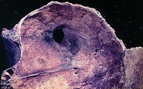
Diagnosis: Case 48 - Lesion is the result of an arterial venous endocranial aneurysm of probable meningeal origin.
Case 47
Skull from the San Miguel Culture of northern Chile that dates from 1000 AD. There is a soft tissue nodule in the nasal cavity.
Submitted by Dr. Marvin J. Allison
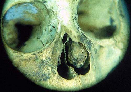
Diagnosis: Case 47 Nasal polyp.
Case 46
Excavation from the island of Guam, Latte phase AD 800-1000. Female: age 40. Images shows bones from 2 thumbs and 5 finger distal phalanges from distal ends. In 2 of the specimens, little remains but the proximal end plate. Four of the proximal end plates appear normal, three are flared and cupped.
Submitted by Bernardo Arriaza and Diane Grembly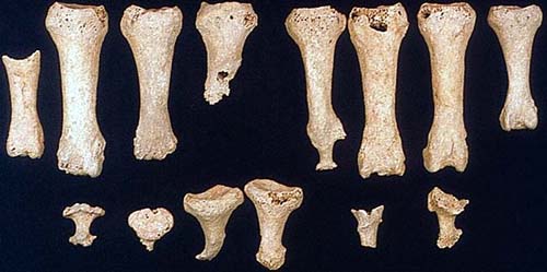
Diagnosis: Case 46 Leprosy.
Case 45
This specimen is from a tamarack bog excavation in Minnesota. The age of this specimen is approximately 8,000 years BP. It is the right mandible of an adult bison (Bison bison).
Submitted by Dr. Standley E. Lewis
Diagnosis: Case 45 Actinomyces bovis. The infected bison jaw appears to represent a bacterial infection, probably caused by the organism Actinomyces bovis.
Case 44
This specimen is from Oman.
Submitted by Dr. Ezio Fulcheri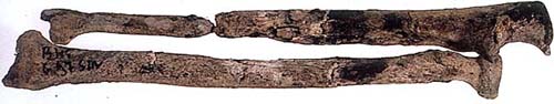
Diagnosis: Case 44 Bone Fracture with diastasis and pseudo-arthrosis.
Associated with mild osteomyelitis in the proximal fragment. This pathology is frequently seen as a complication of severe bone fractures.
Case 43
This bronze figurine from the Cuzco area depicts a medical problem. There was a late Inca custom that, instead of sacrificing a person, a bronze or ceramic effigy was buried with an important man who'd died.
Submitted by Dr. Marvin J. Allison
Diagnosis: Case 43 Potts disease.
Case 42
Two skulls from females approximately 40-years-old; dated 500 AD. Both skulls show severe destruction of bone in the nasal/orbital maxillary area with peripheral extension.
Submitted by Bernardo Arriaza
Diagnosis: Case 42 Infected mucocutaneous leishmaniasis.
Case 41
This specimen is from Tal-Liedna Road in Malta. It is one of the Punic skeletal remains from the 3rd century BC. The specimen is from a young female. The anterior view of the right shoulder joint is shown.
Submitted by S. Ramaswamy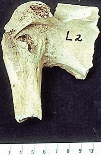
Diagnosis: Case 41 Chronic pyogenic osteomyelitis with ankylosis.
Case 40
A pelvis (above) and a rib (below) of a man (about 32-years-old) from the Diaguita culture that inhabited the area of Chile at the level of La Sirena, spilling over into what is present day Argentina. Their period is approximately 1200 AD-500 BP. They were shepherds, fishermen and farmers of squash, beans and corn.
Submitted by Maria Rosado
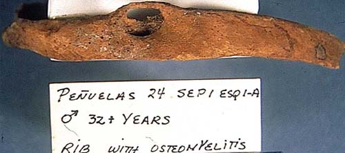
Diagnosis: Case 40--Post-traumatic osteolitis.
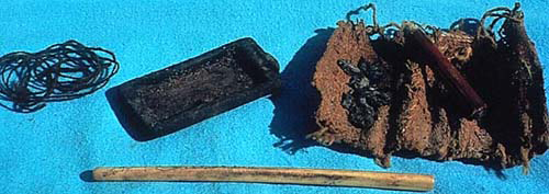
Case 39
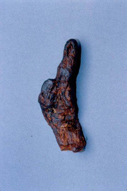
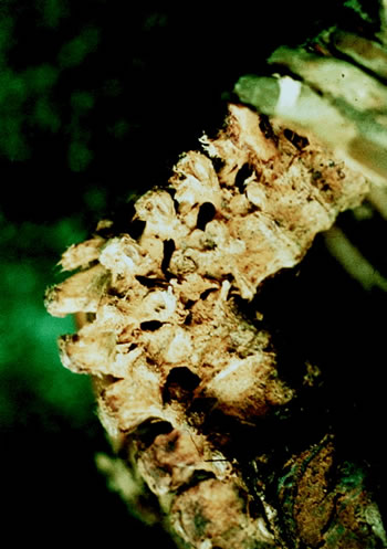
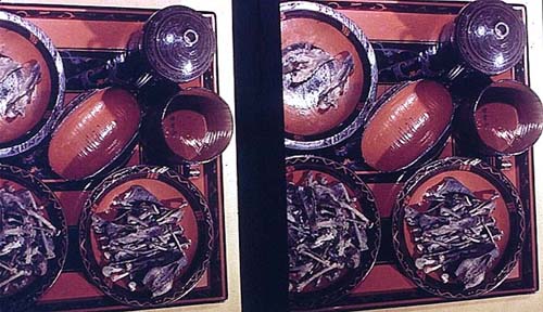
Diagnostic pathology is aided by knowing the society and environment in which the "patient" lived. One of the greatest difficulties in paleopathological diagnosis the general lack of a "clinical history" or even a thorough understanding and knowledge of the times and the society in which the individual under study lived.
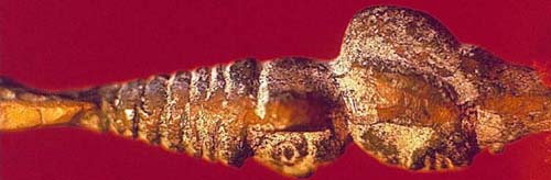
On occasion we do have "diagnostic aids" that are found in the grave along with the body and the slide for this issue is one such aid. This is a kit (all of these items are used to perform a certain ritual or act) from the San Miguel coastal people of Northern Chile. It dates from about 1000 A.D.
1. What are the different items?
2. What would be a possible use of each?
3. What pathology might be expected from frequent usage?
Submitted by: Dr. Marvin J. Allison, Medical College of Virginia, Richmond, Virginia.
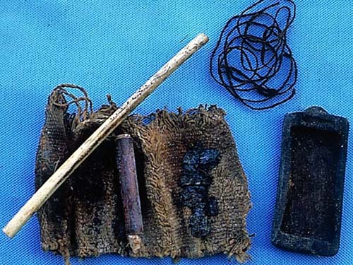
Diagnosis: Case 39 Hallucinogenic Kit.
The use of hallucinogens in South American folk medicine goes back thousands of years. Three major types are still in use today: 1) the San Pedro cactus which is used as a source of mescaline 2) Ayahuasca (Banisteriopsis caapi), a jungle vine which is the source of banisterine 3) Wirca (Piptadenia colubrina), a large, flat seed.
San Pedro is used as a tea in northern Peru. Ayahuasca, as a tea in the Amazon, and Wirca as a powder in southern Peru, Bolivia and Chile. For many years in some graves in northern Chile, archeologists were finding small, wooden trays. Some were plain. Others were carved and decorated with colored stones. It was thought that the trays were used to crush tobacco to make snuff. In recent years, however, complete kits were found along with the seed that was crushed on the tray using a bone spatula.
The powder from the crushed seed would have been brushed into a small pile and aspirated through a bird bone into the nasal mucosa. Powerful hallucinations would have been produced in about twenty minutes. The image shows a kit that is about 1000-years-old. It consists of a wooden tray, seeds of Wirca, a small tube of powder with a cotton stopper, and a bird bone inhaler. All of this would have been wrapped in a cloth and tied with a string. Such kits were generally found in the graves of elderly males.
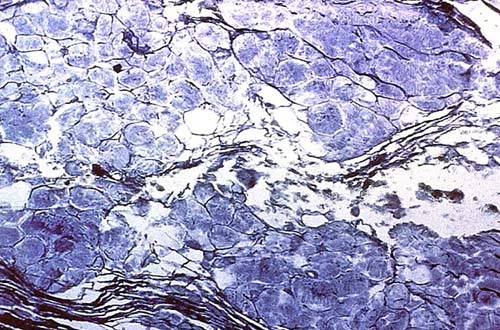
Case 38
A section of the lung from a 3-year-old female, Pre-Columbian mummy from northern Chile.
Submitted by Dr. Jerrold L. Abraham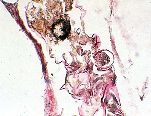
Diagnosis: Case 38 Lung infested by mites.
Case 37
The skull is from an adult male mummy found in the Valle de Lluta, northern Chile, from the Gentilar culture that flourished between 1200 and 1470 AD.
Diagnosis: Case 37 Criba orbitalia.
Case 36
The skull is from a Woodland Indian from the XVI century. It was found in the Tennessee-Virginia border. Although the skull was damaged, the main lesion is clearly visible.
Submitted by Dr. Marvin J. Allison
Diagnosis: Case 36 Posterior palatal cyst.
Reference: Gregg, J. B., Allison, M. J., Clifford, S., Gerszten, E., & Klippel, W. E. (1983). [article title not given]. Journal of the Plains Anthropological Society, 28, p. 293.
Case 35
Mummy of a priest from the early XVII century entombed in the basement of the Iglesia del Carmen Church in Mexico City.
Submitted by Dr. Marvin J. Allison
Diagnosis: Case 35 Smallpox.
Reference: Medicine in Mexico, by Gordon Schendel, University of Texas, 1968
Case 34
Radiograph of the pelvis from a 50-year-old female who was a member of the Maitas Chiribaya culture circa AD 1200. Other osteological findings included lytic lesion in the body of C6 and C7, and an abscess in the posterior intercondyloid fossa of her left tibia.
Submitted by Bernardo Arriaza
Diagnosis: Case 34 Complete bilateral sacro-iliac ankylosis and tumor in the diploe of the left pubic corpus. This is probably a case of seronegative spondyloarthropathy such as ankylosing spondylitis. Incidental finding--a bladder calculus.
Case 33
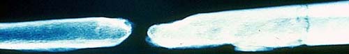
Cross-section of the midshaft of a femur of an Egyptian male, circa 36 BC to 400 AD (the Roman period of Egypt).
Submitted by Megan Cook and C. Anderson, MD
Diagnosis: Case 33 Tetracycline labeling
The slide shows tetracycline labeling similar to that of tetracycline labeled patients under metabolic bone disease investigations. It is postulated that the tetracycline was present in the stored grain.
Case 32
The external auditory canal from a Pre-Columbian mummy (male adult) from Northern Chile (coast).
Submitted by Peter Carlos Gerszten

Diagnosis: Case 32 Exostosis.
Reference: Greg, J. B., & Bass, W. M. (1970). Exostoses in the external auditory canals. Annals of Otology, Rhinology and Laryngology, 79, 4, pp. 834-839.
Case 31
The slide shows the opened gallbladder of an adult, female, Pre-Columbian mummy from Northern Chile.
Submitted by Dr. Marvin J. Allison
Diagnosis: Case 31 Cholecystitis and cholelithiasis.
Reference: [author not given]. (1978, February). Cholelithiasis and cholecystitis in Pre-Columbian Chileans. American Journal of Physical Anthropology, 48, 2, pp. 209-212.
Case 30
The lower portion of the tibias from a Pre-Columbian mummy from Northern Chile.
Submitted by Dr. Marvin J. Allison
Diagnosis: Case 30 Squatting facets. Flexing the lower extremities for extended periods of time produces characteristic impressions on the bones of the joints.
Reference: Ubelaker, D. H. (1978). Human skeletal remains. Illinois: Alidine.
Case 29
Right radius and ulna with proximal fusion from protohistoric adult Indian (Chumash), Santa Rosa Island, CA. The union starts at the radial head and extends distally for a distance of 4.8 cm resulting in 90 degree pronation of the distal ends.
Submitted by Dr. James M. Tenney
Diagnosis: Case 29 Congenital radio-ulnar synostosis.
Reference: Edmonson, A. S., & Crenshaw, A. H. (1980). Campbell's operative orthopaedics. St. Louis, MO: C. V. Mosby.
Case 28
Tibias of a Roman soldier (50-60-years old) from Gerulata I burial ground dated from the 3rd-4th century AC (from the Slovak National Museum, Bratislava).
Submitted by Dr. Václav Smrcka, Dedická
Diagnosis: Case 28 After fractures of the distal third of each tibia, the slide shows a callous formation on the right with union of the fibula. On the left side an osteomyelitis is shown.
Case 27
A skin lesion of an Egyptian mummy taken from the Marro Collection, Turin, Italy (Cat. No. 61). The lesion is from the neck, and it is presented as a raised, hard, roundish mass, brown in color, (0.7 cm in diameter).
Submitted by Dr. Ezio Fulcheri
Diagnosis: Case 27 Verrucous lesion.
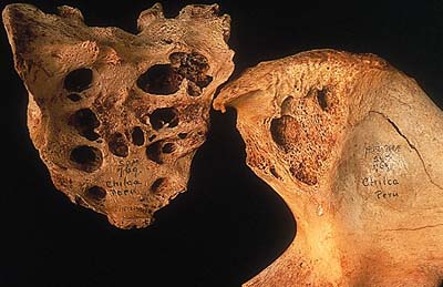
Case 26
Picture shows the posterior surface of two intentionally deformed skulls excavated from Pre-Columbian sites in Northern Chile.
Submitted by Peter C. Gerszten
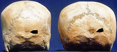
Diagnosis: Case 26 Necrosis of the occipital bones resulting from the pressure of a head deforming apparatus. The left skull also has an intercalary bone (wormian bone).
Case 25
Infant male mummy heart from the Anasazi culture (Canyon de Chelly, northern Arizona, about 600 AD ± 95 years). The illustration shows the outflow tract of the left ventricle, the interventricular septum, and valvular cusps. Other autopsy findings included cardiomegaly and peripheral edema, including ascites.
Submitted by Dr. Abel L. Robertson
Diagnosis: Case 25 Common atrial ventricular canal.
Case 24
Adult male mummy from the Cabuza culture (Northern Chile about 450 AD). This picture shows the opened chest with the right lung exposed in more detail than the left. The autopsy findings included right ventricular dilatation and massive gastric dilatation.
Submitted by Dr. Arthur C. Aufderheide
Diagnosis: Case 24 Diffuse bilateral bullows emphysema.
Case 23
H&E section of one of eight skin lesions on the lower extremities of an frozen Incan mummy (9-year-old) found at high altitude in the Andes.
Submitted by Patrick Horne
Diagnosis: Case 23 Microscopic section showed a rare childhood tumor of skin--angiokeratoma circumscriptum.
Reference: (1984) Bulletin NY Academy of Medicine, 60, 9, pp. 925-931
Case 22
Autopsy from a frozen mummy; microscopic section of the lung.
Submitted by Dr. Michael R. Zimmerman
Diagnosis: Case 22 Anthracosis and fibrosis.
Case 21
Autopsy of an ancient cadaver from the Han Dynasty (207 BC-220 AD)--the tomb was found in Hunan Province. Above are relics found in the tomb. Below is a section of the liver.
Submitted by Dr. Longxiang Peng

Diagnosis: Case 21 Schistosome
Section of the liver shows a nodule containing several schistosome eggs. Among the valuable relics there are many lacquers. Shown is some of the dinner set.
Case 20
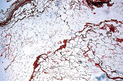
This low-power photomicrograph is of breast tissue from a frozen mummy of a 42-year-old Eskimo woman who died in northern Alaska approximately 500 years ago. Both breasts were prominent on gross examination.
Submitted by Dr. Michael Zimmerman
Diagnosis: Case 20 Pregnant breast.
Case 19
The left humerus was the only material recovered from this adult. The length is 161 mm. The epiphysis are well-fused. The proximal shaft is thickened and robust with enlarged greater and lesser tubercles and an extremely prominent deltoid tuberosity. The olecranon fossa is small, as are the radial and coronoid fossae. The medial epicondyle is exaggerated, giving a flared appearance distally.
Submitted by Dr. James M. Tenney
Diagnosis: Case 19 Achondroplastic dwarf.
Source of material: Lowie Museum of Anthropology, Berkeley, CA, Accession No. 12-6616)
References:
Hoffman, J. M. (1976). Contributions of the University of California Archeological Research Facility, 30, pp. 65-119.
Case 18
Right and left humerus, ulna and radius of a 22-24-year-old male from 350 AD.
Measurements (mm)
Right Left
Humerus 305 235
Ulna 244 268
Radius 199 252
The glenoid cavity of the left scapula was eroded and its shape deformed. The soft tissues appeared normal.
Diagnosis: Case 18 Possible Accident
X-ray studies suggest that the lesion of the left humerus and right forearm are related and caused by an accident. The left humerus is a fracture resulting in remodeling of the bone with subsequent loss of scapula joint contact and/or formation of a new articulation. The right forearm can be classified as a reverse Madelung deformity.
Case 17
Skin biopsy from a woman approximately 40-years-old from 350 AD. The section was taken from her forearm. It represents one of numerous 1-2 mm papular, rough-surfaced, elevated lesions. For orientation, the epidermis is missing, but the papillary and reticular dermis may be distinguished.
Submitted by Dr. H. Sasken
Diagnosis: Case 17 Systemic vasculitis (trepanematosis).
Case 16
This picture shows the gross examination of the thoracic cavity of an 11-year-old male mummy from 350 AD.
Diagnosis: Case 16 Bilateral emphysema.
Case 15
Mummy of a 40-year-old man of the Huari culture. The picture shows the base of the skull.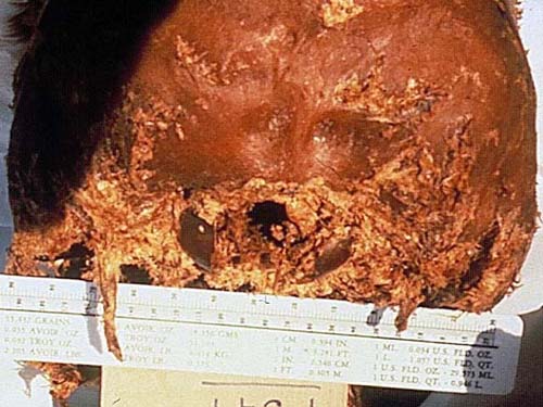
Diagnosis: Case 15 Elongated styloid process (Eagle's Syndrome).
Reference: [No author given] (1980, January). Elongated styloid process in a Pre-Columbian Peruvian. Journal of Dental Research, p. 79.
Case 14
Mummy of a 14-year-old boy. There is a mass that measures 4x4x2 cm high 6 cm below the axilla, on the right side of the nipple. Included is an image of a microscopic section from the tumor mass--H&E.
Diagnosis: Case 14 Lipoma.
Case 13
Male, age 25 dating from 250 AD. The external examination showed a circular wound in the neck into the trachea and a large, 20 cm wound in the left abdomen with the left hand holding loops of the intestine. The head had been cut off and was probably taken as a trophy. Organs were present and normal except for one kidney.
Diagnosis: Case 13 Hydronephrosis of right kidney caused by a calculus lodged in the right ureter.
Case 12
Mummy liver section from a 5-year-old Inca child. On gross examination, the liver was enlarged and the microscopic lesions seen were present throughout the liver parenchyma.
Diagnosis: Case 12 Echinococcosis (E. multilocularis)
Multilocular cysts are found in this species with the pox, and possibly puma, as the definitive hosts.
Case 11
A newborn child from a cemetery in Ica, Peru shows occipital bone lesions and similar lesions in the parietal bones.
Diagnosis: Case 11 Lückenschädel Syndrome.
Case 10
Adult with skin lesions and skull with "saber shin" tibias from an Atacameña, Chilean mummy dating around 290 AD.
Diagnosis: Case 10 Trepanematosis.
Case 09
This skull is from a 45-50-year-old woman from the Maitas-Chiribaya culture who died around 1000 AD. The cemetery was in a ceremonial site and numerous dogs were found in the excavation, some also with ceremonial burials. The lesion seen in the slide is a solitary bone lesion involving the nose, right eye, left frontal sinus, right maxillary sinus and posterior palate.
Diagnosis: Case 09 Chronic leishmaniasis with a superimposed infection.
Case 08
This mummy of an 8-year-old male dates from 300 AD. A review of the internal organs shows the following findings:
-
the heart (enclosed picture)
- the lung (pneumonia) and the liver (multiple abscesses)
Diagnosis: Case 08 Cardiomegaly and fibrous and fibrinous pericarditis, a complication of a fatal pneumonia.
Case 07
Male mummy (17-19 years old) dating about 300 AD. Review of the organs showed the lungs with emphysematous changes. The only other abnormality is in the kidneys. Cross-section is shown in the picture. The appearance is similar in both kidneys.
Diagnosis: Case 07 Polycystic Kidney Disease
The kidney showed gross and microscopic features of this disease.
Case 06
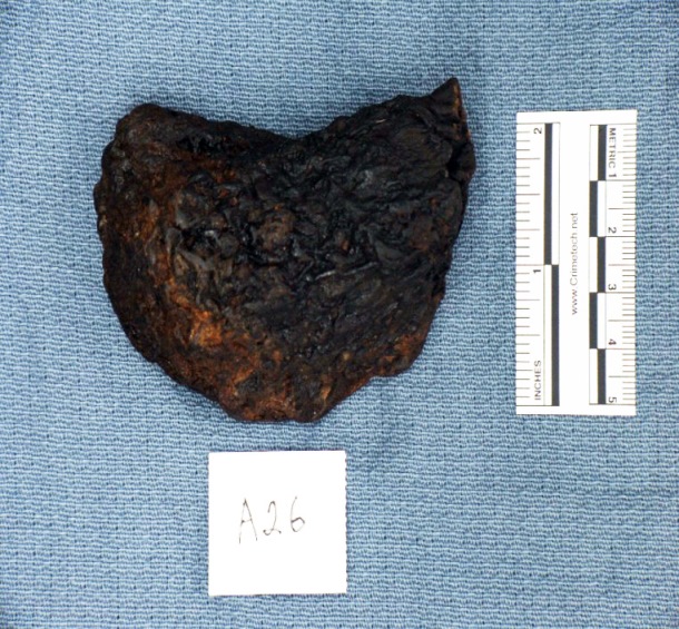
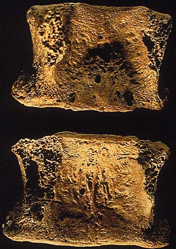
A male mummy, over 40 years old, from 2800 BP was autopsied. On opening the intact thoracic cavity, a hard, stony structure was found in the mediastinum. It measured 40x18 mm, was greenish in color and was partially covered by the pleura. Microscopically, there was no reaction around the structure.
Diagnosis: Case 06 Projectile Point still in the mediastinum. The skin was completely healed. The time of the injury was long before death.
Case 05
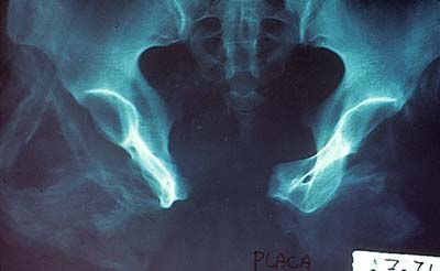
A mummy bundle with the skeleton of a 45-year-old female from 800 AD was salvaged near Arica, Chile. At autopsy, nine bones had gross evidence of pathology: the skull, the right and left innominate, the right femur, sacrum, the eighth thoracic vertebra, the fourth and fifth lumbar vertebrae, and the sternum.
The skull had three lesions of the parietal bones near the sagittal suture. The largest was a lytic lesion that began in the diploe and perforated the inner and outer tables equally, leaving a ragged opening 35x30 mm. To the right of this opening was a nonperforating lesion 17x16 mm and to the left a smaller nonperforating lesion 15x15 mm. The nonperforating lesions were seen as a roughening and incipient crumbling of the outer table. Radiographically, the nonperforating lesions were considered to be sclerotic lesions. There was a third non-perforating lesion about 10x10 mm in the central portion of the occipital bone.
Both innominate bones were extensively involved in the disease, seen in second image. The right femur had a lytic lesion at the proximal end which involved the neck. There were three lesions of the vertebrae. The entire eighth thoracic body was destroyed. The fourth and fifth lumbar each showed a small incipient lesion in the body. There was a small lytic lesion in the sternum at the level of costal notch II.
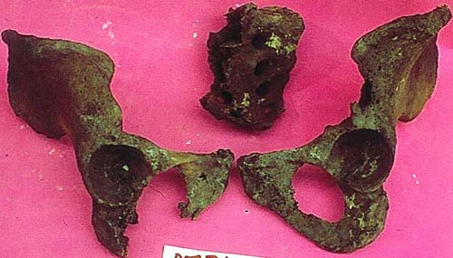
Diagnosis: Case 05 Metastatic Tumor, most likely from a primary of the breast.
Reference: Bulletin of the New York Academy of Medicine, 56, 585-587, 1980
Case 04
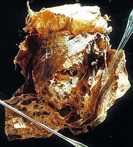
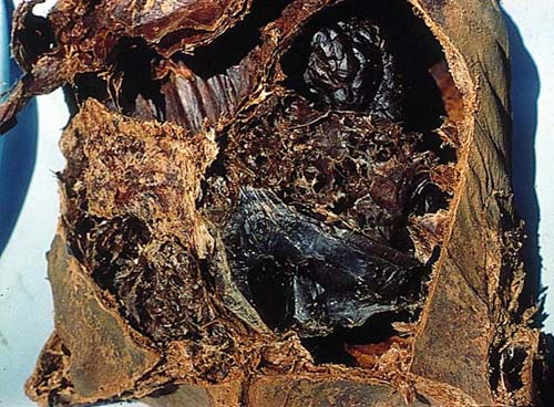
Image A. Mummy of a 56-year-old adult female, buried in the Azapa Valley, Chile, who died around 290 AD. Aside from the usual grave goods associated with mummies in this time period, there were large numbers of tropical bird feathers. On autopsy:
- The right lung on the lateral surface was firmly adherent to the costal pleura at the level of the 3-5th ribs.
- Separation of the adhesion revealed a large cavity 95x70x55mm (slide 1). The cavity contained a blackish granular material.
- Right tracheobronchial lymph nodes were the size of marbles.
- Three sets of enlarged mediastinal nodes were noted.
- The right kidney was thickened and irregular in shape when compared to the left.
- Histological sections of the right lung cavity wall revealed numerous red blood cells and yeast-like organisms 6-12 microns.
Image B. EM studies revealed the organisms seen in image B in the form of a "ship's wheel".
IMAGE A
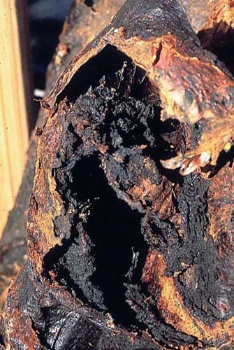
IMAGE B
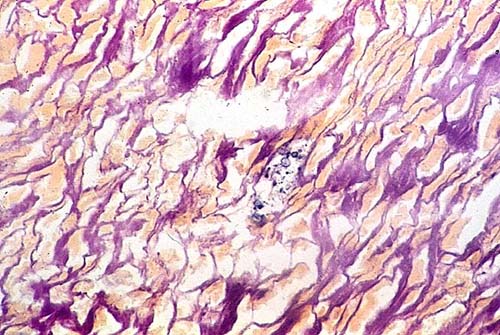
Diagnosis: Case 04 South American blastomycosis.
Case 03
This skull is from San Pedro de Atacama, Chile dating from approximately the 3rd century AD. The two photographs are representative of what is a very chronic process that did not involve the internal table of the skull in any way, although it probably limited the use of the eye on that side--due to compression, not direct invasion.
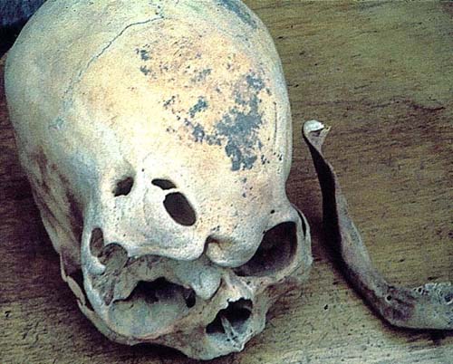
Diagnosis: Case 03 Sinus mucocele.
Case 02
The skull was from a woman around 35-40 years old. It was salvaged from "Falda de Morro" cemetery in Arica, Chile. Visual and X-ray revealed a single, lytic lesion at the junction of the parietal bones with the occipital. It measures 50x45 mm with short, bony spicules. The inner table hole was larger than the outer.
Diagnosis: Case 02 Unifocal eosinophilic granuloma.
Case 01
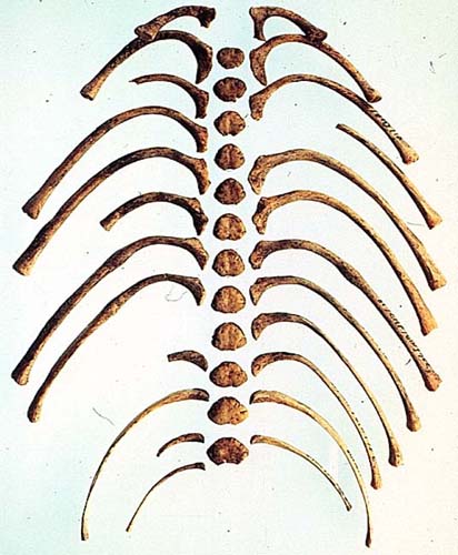
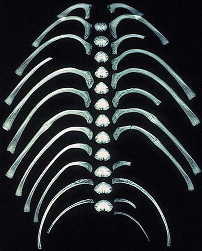
Four bones from the single skeleton of a person who was about 30-years-old were recovered from a vandalized Huari cemetery in the Ingenio Valley, Ica, Peru (Huari culture 800-1200 AD). All bones were heavier than normal (a normal femur weighs 350 gms). A radiograph showed a 2/3 reduction of the marrow cavity. A microscopic of the rib is shown with multiple thickening of the cortical area.

Diagnosis: Case 01 Primary generalized hyperostosis.
Reference: This case was published in the 1976 MCV Quarterly, 12, 2, pp.49-51.